Hemolyzed Blood: What Hemolysis Means for Your Research Samples
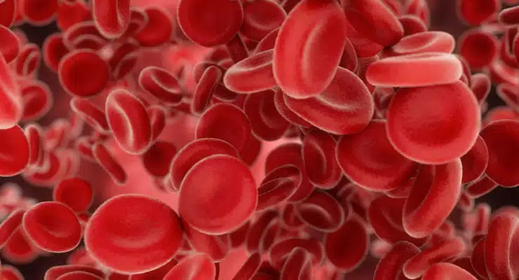
Hemolyzed blood is blood in which red blood cells have ruptured and released their contents into the surrounding fluid. Hemolysis — the rupture of red blood cells — can happen at any point, both in vivo (inside the body) and in vitro (after a sample is collected), and it is one of the most common reasons a blood sample is rejected for research or diagnostic use.
What Does Hemolyzed Mean?
When blood is described as hemolyzed, it means the membranes of the red blood cells have broken down and hemoglobin and other intracellular components have leaked into the serum or plasma. A normal serum or plasma sample is pale yellow; a hemolyzed blood sample takes on a pink-to-red tint that deepens with the degree of hemolysis.
The distinction matters because the released contents — hemoglobin, potassium, enzymes, and proteases — change the biochemistry of the sample and can invalidate downstream measurements. Understanding what hemolyzed means, what causes it, and how it is graded is essential for anyone sourcing or working with human blood samples.
How Hemolysis Affects Lab Results
Hemolysis can significantly skew study and diagnostic results. Its best-documented effect is on potassium measurements, because red blood cells are potassium-rich and release it when they rupture. It also distorts measurements of enzymes and analytes including:
- Lactate dehydrogenase (LDH) and aspartate aminotransferase (AST) — elevated by intracellular release
- Potassium — falsely elevated (pseudohyperkalemia)
- Insulin and cardiac troponin — degraded by proteases released from ruptured cells
- Spectrophotometric assays — interfered with by free hemoglobin’s color and absorbance
Because free hemoglobin also lowers haptoglobin and can raise bilirubin, hemolysis can mimic or mask real biological signals — a serious problem for biomarker discovery and clinical research.
What Causes Hemolyzed Blood? In Vitro vs. In Vivo
There are two broad categories of hemolysis, and telling them apart matters for interpreting a sample.
In Vitro Hemolysis (During and After Collection)
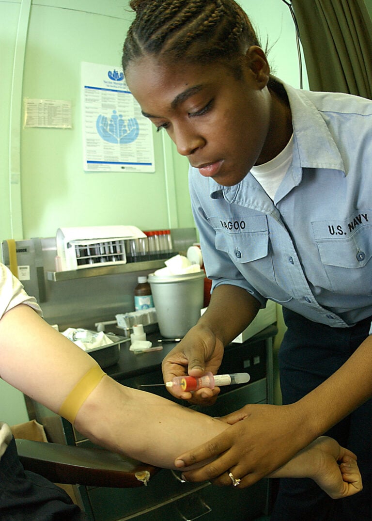
Most sample hemolysis — an estimated 50–75% of cases — happens at the moment blood is drawn. Common causes include:
- Using the wrong gauge needle or applying too much suction
- Leaving the tourniquet on too long
- Shaking or mixing the tube too vigorously
- A difficult or traumatic draw
- Improper transportation, temperature excursions, or prolonged storage
This is why phlebotomy technique and sample handling are the highest-leverage places to prevent hemolysis.
In Vivo Hemolysis (Inside the Body)
When hemolysis occurs in vivo, red blood cells die prematurely in circulation. Potential causes include:
- Antigen–antibody (immune) reactions
- Chemical reactions and toxins
- Hemolytic anemias such as sickle cell anemia or thalassemia
- Mechanical interventions such as dialysis or a heart–lung bypass machine
- Artificial heart valves
The Hemolysis Index: Grades of Hemolysis
A sample isn’t simply “hemolyzed” or “not.” Hemolysis falls on a continuous scale — often expressed as a hemolysis index — from none to gross hemolysis. Sanguine guidelines dictate that samples are discarded when hemolysis meets or exceeds 200 mg/dL of free hemoglobin.
How the Color of a Sample Reveals Hemolysis
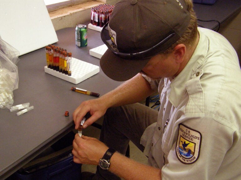
After a sample is spun, its color indicates the degree of hemolysis. A clean sample ranges from pale yellow (no hemolysis) toward pink and then dark red as hemolysis increases. The redder the shade, the more hemolyzed the sample — a fast visual screen used alongside quantitative measurement.
How to Prevent Hemolysis
Because the large majority of hemolysis happens at draw, prevention starts with the phlebotomist. The most effective steps are:
- Highly trained, carefully vetted staff who understand and avoid the causes of hemolysis
- Correct needle gauge, controlled suction, and gentle mixing
- Careful, cushioned transport within the proper temperature range
- Never freezing samples that are intended to be separated
“Although the root cause of hemolysis is difficult to determine on a case-by-case basis, Sanguine does its best to mitigate the risks that can lead to hemolysis to ensure scientists receive specimens that are useful for their research,” says Sagar Amin, CCRA, Biorepository Project Manager at Sanguine.
Frequently Asked Questions
What does hemolyzed mean?
Hemolyzed means red blood cells in a sample have ruptured, releasing hemoglobin and other cell contents into the serum or plasma and giving the sample a pink-to-red tint.
What causes hemolyzed blood?
Most often it’s the blood draw itself (wrong needle, too much suction, vigorous shaking, prolonged tourniquet) or poor handling and storage. It can also occur in vivo from immune reactions, toxins, hemolytic anemias, or mechanical devices.
Why is hemolyzed blood a problem for research?
Hemolysis skews analytes such as potassium, LDH, and AST, degrades proteins like insulin and troponin, and interferes with spectrophotometric assays — so a hemolyzed sample can produce misleading results.
How is the degree of hemolysis measured?
By a hemolysis index (free hemoglobin concentration) and visual grading of sample color after centrifugation. Sanguine discards samples at or above 200 mg/dL.
Related Resources
Hemolysis is a key quality consideration when sourcing human serum, human plasma, and human whole blood for research — each is screened for hemolysis before release. For help choosing between matrices, see our guide to serum vs. plasma.