Epithelial to Mesenchymal Transition: the Key to Cancer Metastasis
Picture a science-fiction invasion story: before the main force arrives, a single scout is sent ahead, built to survive a harsh journey, to find fertile new ground.
Once the scout succeeds, the full invasion follows. Nature uses this same strategy — a hardy virus particle or a sporulated bacterium survives harsh conditions just long enough to reach a new host, then takes over once it arrives.
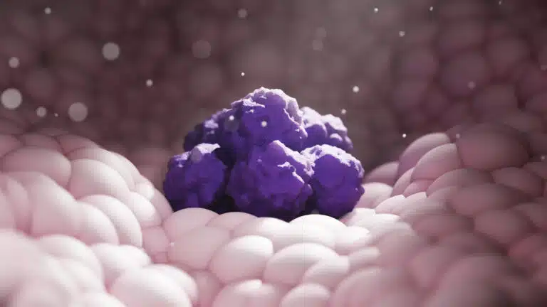
Cancer metastasis works the same way. Cancer cells break away from the primary tumor, survive a harsh journey through the body, and form new colonies in other tissues. These metastases are dangerous. In some cancers, like melanoma, it’s specifically the spread to vital organs such as the brain that makes the disease life-threatening.
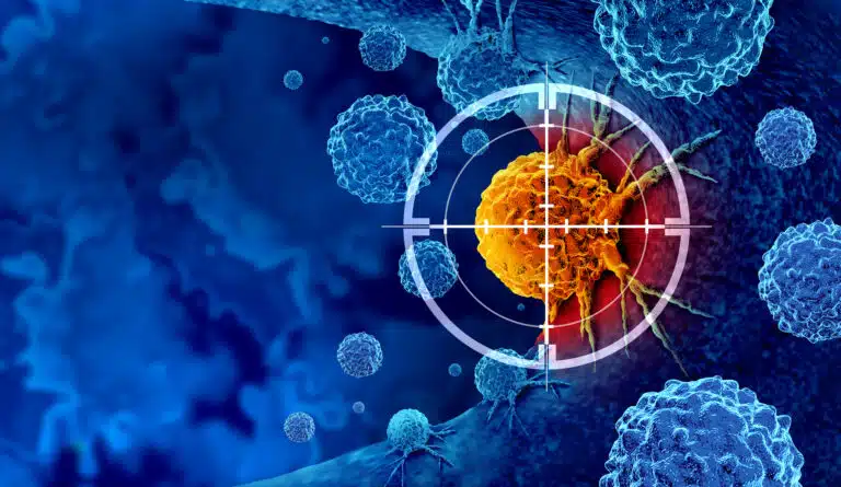
One of the biggest questions in cancer biology is how metastasis actually happens — and a central piece of the answer is the epithelial to mesenchymal transition (EMT). EMT is what turns a cancer cell into a "scout": able to invade blood vessels, survive the journey to a new site, and resist most therapies wherever it ends up.
EMT isn’t unique to cancer. In normal development, it’s a reversible process used in embryonic gastrulation1 and cardiac development1,2. In adults, it’s involved in wound healing3.
But EMT also drives harmful processes — fibrosis of injured tissue1, and cancer development and progression1-4. This article focuses on EMT’s role in tumorigenesis, cancer growth, treatment resistance, and metastasis.
How EMT Drives Cancer Metastasis
EMT happens in many solid tumors of epithelial origin. Most of these tumors are carcinomas, which are normally kept in place by the basement membrane1. But once a cancer cell takes on mesenchymal traits, it can move — infiltrating blood and lymphatic vessels and eventually spreading to new sites1,5.
As a cancer progresses, two things happen together:
- Epithelial features go down — including the adhesion marker E-cadherin3 and adhesion components like tight junctions, cytokeratins, and desmosomes5.
- Mesenchymal markers go up — including N-Cadherin, Vimentin, and Fibronectin3.
Several mechanisms drive this shift together: epigenetic changes, post-translational modifications of proteins, transcriptional silencing by noncoding RNAs (ncRNAs), and activation of epithelial-to-mesenchymal transcription factors (EMT-TFs)3. All of these mechanisms matter, but this article focuses on the main drivers: the EMT-TFs.
The SNAIL Family
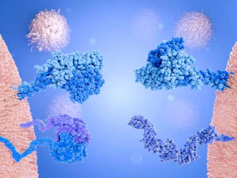
Many transcription factors act as EMT-TFs, but most fall into three families: SNAIL, ZEB, and TWIST. SNAIL itself has three members: SNAIL (Snail1), SLUG (Snail2), and SMUC (Snail3)5. These proteins are zinc finger nucleases, and they sit at the center of the EMT phenotype.
- SNAIL — and possibly SLUG — directly represses E-cadherin by binding its promoter, CDH13.
- The SNAIL family also represses desmoplakin, adherens junctions, occludins, and cytokeratin once activated5.
Little is known about SMUC’s role in normal or diseased development. But SNAIL, unlike SLUG, is so central to metastasis that it’s been proposed as an independent prognostic marker for how aggressive and metastatic a cancer will be5.
The SNAIL family doesn’t just drive EMT directly — it also upregulates other EMT-TFs, like ZEB1 and ZEB2, spreading the EMT program further5.
The ZEB Family
The Zinc-finger E-box-binding homeobox (ZEB) family are also zinc-finger nuclease transcription factors3. Like SNAIL, ZEB1 and ZEB2 bind the E-cadherin promoter5. They also:
- Downregulate tight/gap junctions, desmosomes, and markers of cell polarity5.
- Repress P- and R-cadherins, two other markers that normally limit epithelial cell movement5.
ZEB1 usually isn’t found on non-cancerous cells, but shows up strongly on many cancer types5. ZEB2, in contrast, is present on normal epithelial cells too — but is much more upregulated in cancer cells5.
The TWIST Family
The last group is TWIST, a basic helix-loop-helix transcription factor involved in several steps of embryonic development1. TWISTs are essential for embryogenesis, but absent in normal adult epithelium5. As a cancer cell progresses, TWIST1 and TWIST2 appear, and their activity rises alongside tumor progression5. TWIST1 helps regulate some of SLUG’s EMT effects and directly drives N-cadherin expression — but it isn’t directly linked to E-cadherin downregulation5.
EMT-TFs in Tumor Invasion and Metastasis
EMT-TFs drive tumorigenesis and tumor progression by suppressing senescence and boosting cell cycle proliferation5. On their own, though, EMT-TFs aren’t enough to cause tumorigenesis — they need another triggering event3. So EMT-TFs likely act as facilitators of tumorigenesis, not tumorigenic factors on their own.
EMT is also key to tumor invasion and metastasis. Beyond the adhesion molecule changes already mentioned, activating EMT-TFs raises levels of matrix metalloproteinases (MMPs) — enzymes that break down the extracellular matrix and let cancer cells invade3. The SNAIL family activates MMP1, MMP2, MMP7, and MT1-MP 5.
In turn, higher MMP levels further activate EMT-TFs, creating a feed-forward loop3. EMT can drive invasion beyond matrix degradation too: TWIST1 helps form invadopodia, structures linked to invasiveness3.
The Tumor Microenvironment’s Role in EMT
Metastasis doesn’t just come from changes inside the cancer cell — changes in the host tissue and the target microenvironment matter too4. For example, TGF-β normally acts as a tumor suppressor, but can actually enhance invasion in later-stage tumors2. TGF-β activates both the SNAIL and TWIST families of EMT-TFs2,5.
TGF-β itself may come from myeloid derived suppressor cells and CD11b+/F4/80+ tumor-associated macrophages (TAMs) in the primary tumor, which keeps the EMT phenotype going4. TAMs also release fibroblast growth factor (FGF), epidermal growth factor (EGF), and macrophage colony stimulating factor (CSF-1), which support EMT-driven invasion and recruit immune cells into a microenvironment that favors metastasis4.
TNF-α, usually linked to anti-tumor activity, also stabilizes SNAIL expression — putting it, too, in EMT’s corner3. Immune cells aren’t the only stromal players. Mesenchymal stem cells (MSCs) and cancer-associated fibroblasts (CAFs) also help start and spread EMT4.
EMT and Therapy Resistance
One of the most clinically important effects of EMT is drug resistance:
- Higher ZEB1 levels correlate with doxorubicin resistance in breast cancer5, and both ZEB1 and ZEB2 protect cancer cells against cisplatin therapy5.
- TWIST1 causes resistance to paclitaxel. TWIST1 and TWIST2 also block daunorubicin, by preventing degradation of the anti-apoptotic protein Bcl-2, in bladder, ovarian, and prostate cancer5.
EMT has also been shown to give cells a cancer stem cell-like phenotype2,4 — a phenotype also known for resisting therapy5. TGF-β-driven EMT activates stem cell proteins like Sox2, PDGFB, and LIF 2, and ZEB1 helps form and maintain the stem cell phenotype in some cancers5.
Still, activating EMT isn’t required to get a stem cell-like phenotype3 — EMT-TFs aren’t always involved in dedifferentiation3. The evidence is actually mixed: colorectal cancer spheroids show higher SNAIL levels, but overexpressing SNAIL and SLUG in ovarian cancer pushes those cancers away from a stem cell phenotype5.
Challenges in Studying EMT
Beyond the treatment challenges EMT creates, one of the biggest obstacles in the field is simply identifying EMT/MET in cancer in vivo4. Because EMT results from an interplay between the tumor and its microenvironment, researchers haven’t been able to definitively prove EMT’s role beyond the stromal epithelium4. The field needs better markers to tell tumor epithelial cells apart from normal ones, and better ways to trace human cancer lineages in vivo4.
There’s a similar problem with MET — the reverse of EMT, and the end result of metastasis. So far, solid evidence for MET exists only in in vitro studies and xenograft experiments4. MET explains why metastatic tumors resemble the primary tumor’s phenotype, but it doesn’t explain the underlying system4.
Researchers have proposed better methods for studying EMT, like intravital 2-photon microscopy4. But EMT events happen sporadically, which makes them hard to observe even with this technology4. For now, researchers rely on spontaneous tumor-forming mouse models, or xenograft models using immortalized, highly metastatic cancer lines.
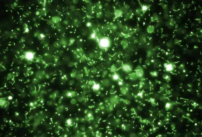
There’s also no anatomical way to tell mesenchymal and epithelial cells apart just by looking. One proposed fix: build tumor lines that express reporter genes linked to promoters for epithelial or mesenchymal cell fates4.
Combining an intravital two-photon system with a xenograft of a highly metastatic cancer line carrying these mesenchymal/epithelial reporter constructs would be the most workable model for studying EMT in real time. As technology improves at tracking individual cell populations in real time, researchers should be able to nail down the mechanism of EMT in metastasis with more confidence.
References:
1 Lim, J. & Thiery, J. P. Epithelial-mesenchymal transitions: insights from development. Development 139, 3471-3486, doi:10.1242/dev.071209 (2012).
2 Massagué, J. TGFβ signalling in context. Nat Rev Mol Cell Biol 13, 616-630, doi:10.1038/nrm3434 (2012).
3 Craene, B. D. & Berx, G. Regulatory networks defining EMT during cancer initiation and progression. Nat Rev Cancer 13, 97-110, doi:10.1038/nrc3447 (2012).
4 Gao, D., Vahdat, L. T., Wong, S., Chang, J. C. & Mittal, V. Microenvironmental regulation of epithelial-mesenchymal transitions in cancer. Cancer Res 72, 4883-4889, doi:10.1158/0008-5472.can-12-1223 (2012).
5 Sánchez-Tilló, E. et al. EMT-activating transcription factors in cancer: beyond EMT and tumor invasiveness. Cell Mol Life Sci 69, 3429-3456, doi:10.1007/s00018-012-1122-2 (2012).