CD4+ T Cells in the Tumor Microenvironment: Allies, Adversaries, and Everything In Between
The tumor microenvironment (TME) is a battleground. Immune cells try to eliminate cancer, while tumors use many strategies to survive. Among the immune cells in this fight, CD4+ T cells play a uniquely complex role. Depending on their subset and the signals they get inside the TME, CD4+ helper T cells can either drive strong anti-tumor immune responses or actively suppress them. Understanding this duality — and the molecular mechanisms that tip the balance either way — is one of the most important questions in cancer immunotherapy research today. This article covers what researchers need to know about CD4+ T cell biology in tumors, and why this complexity makes primary cell quality more important than ever.
Why CD4+ T Cells Matter in the TME
For much of the history of cancer immunology, CD8+ cytotoxic T cells got the spotlight as the main anti-tumor effectors, with CD4+ T cells cast as supporting players. That view has changed. We now know CD4+ T cells do several jobs in anti-tumor immunity that CD8+ T cells can’t do on their own:
- They license dendritic cells to prime CD8+ T cells effectively
- They sustain CD8+ T cell effector function and memory formation during chronic antigen exposure
- They coordinate macrophage activation, B cell responses, and NK cell recruitment within the tumor stroma
- In certain contexts — particularly tumors that downregulate MHC Class I to evade immunity — activated CD4+ T cells can directly kill tumor cells. They do this through perforin/granzyme and Fas/FasL mechanisms, independent of CD8+ T cells
But the TME also contains a distinct population of CD4+ T cells working in the opposite direction: regulatory T cells (Tregs). Tregs actively suppress anti-tumor immunity and help tumors evade clearance. The net effect of CD4+ T cell infiltration in any given tumor depends heavily on the ratio and functional state of these competing populations.
Pro-Tumor vs. Anti-Tumor CD4+ T Cell Subsets in the TME
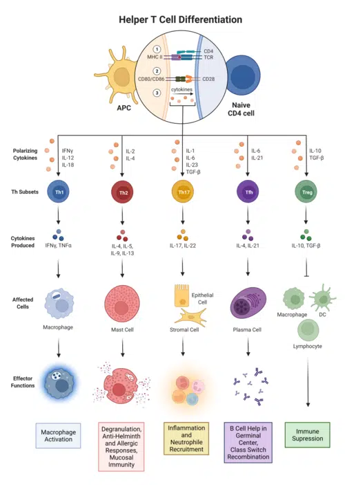
The same flexibility that makes CD4+ helper T cells so versatile in normal immune responses becomes a double-edged sword in tumors. The cytokine environment inside the TME shapes which subset naive and memory CD4+ T cells become when they encounter tumor antigens.
Th1 Cells: The Anti-Tumor Effectors
Th1-polarized CD4+ T cells are the main pro-inflammatory, anti-tumor subset. Driven by IL-12 and IFN-γ signaling and marked by T-bet expression, Th1 cells produce high levels of IFN-γ and TNF-α. This activates macrophages, boosts MHC Class I expression on tumor cells, promotes cross-presentation by dendritic cells, and sustains CD8+ cytotoxic T cell function within the tumor. High intratumoral Th1 infiltration consistently tracks with better prognosis across multiple solid tumor types, including colorectal, breast, ovarian, and lung cancers. Th1 cytokine signatures are among the most reliable transcriptomic predictors of response to immune checkpoint inhibitors.
Regulatory T Cells (Tregs): The Immunosuppressive Counterforce
Foxp3+ Tregs are the best-characterized immunosuppressive CD4+ T cell population in the TME. They build up in tumors through several mechanisms. Tumor cells and tumor-associated macrophages actively recruit them via CCL22 and CCL17 chemokines. Tumor-derived TGF-β and IL-10 drive their preferential expansion. And conventional CD4+ T cells convert into induced Tregs within the suppressive TME.
Once established in the tumor, Tregs suppress anti-tumor immunity through several mechanisms:
- Secreting TGF-β, IL-10, and IL-35 to directly inhibit effector T cells
- Consuming IL-2, which starves CD8+ T cells of a critical survival signal
- CTLA-4-mediated downregulation of B7 co-stimulatory molecules on dendritic cells
- Direct contact-dependent suppression of natural killer cells
High intratumoral Treg infiltration tracks with poor prognosis in most solid tumor types. The ratio of effector CD4+ T cells to Tregs is a meaningful predictor of both spontaneous and therapy-driven tumor control.
Th17 Cells: Context-Dependent and Contested
The role of Th17 cells in tumor immunity is genuinely contested and seems to depend on tumor type. In some settings — particularly colorectal cancer — Th17 infiltration tracks with better outcomes, possibly through IL-17-driven recruitment of anti-tumor innate immune cells or through Th17-to-Th1 plasticity. In others, Th17 cells have been linked to pro-tumor inflammation, angiogenesis, and resistance to checkpoint blockade. For researchers modeling Th17 biology in the TME, this context-dependence raises the stakes for starting material quality and experimental design. That includes the cytokine conditions used to polarize primary human CD4+ T cells.
T Follicular Helper (Tfh) Cells and Tertiary Lymphoid Structures
Tfh cells are a specialized CD4+ subset that normally supports germinal center reactions in lymph nodes. They have become an important part of the anti-tumor immune response through their role in forming and sustaining tertiary lymphoid structures (TLS) within tumors. TLS are ectopic lymphoid clusters that develop in the tumor stroma. They’re now recognized as strong positive prognostic indicators across multiple cancer types, linked to better survival and stronger response to immunotherapy. Tfh-mediated support of B cells within TLS drives local antibody production and boosts cytotoxic T cell priming, creating an intratumoral immune hub that works much like a lymph node.
SanguineBio’s human CD4+ T cells are isolated from healthy, screened donors and characterized for suppression experiments and TME co-culture models.
How Tumors Hijack CD4+ T Cell Biology
Tumors aren’t passive targets. They actively reshape the cytokine environment to steer infiltrating CD4+ T cells away from anti-tumor effector fates and toward immunosuppressive ones. Researchers have described several mechanisms in detail:
TGF-β secretion is one of the most potent tumor immune-evasion strategies. Tumor-derived TGF-β suppresses Th1 differentiation, promotes Treg conversion from conventional CD4+ T cells, and blocks the cytotoxic function of both CD4+ and CD8+ effector populations. It also promotes Th17 differentiation when IL-6 is present, and the resulting Th17 cells can add to immunosuppressive and pro-angiogenic signaling in the tumor stroma.
IDO1 (indoleamine 2,3-dioxygenase 1) is expressed by tumor cells and tolerogenic dendritic cells. It depletes tryptophan — an amino acid essential for T cell activation — and generates kynurenine metabolites that directly promote Treg differentiation and suppress effector T cell function. IDO1 activity creates a local metabolic environment that turns pro-inflammatory CD4+ T cell responses into tolerogenic ones.
Adenosine signaling is generated in the hypoxic tumor core through CD39/CD73 enzyme activity, which is highly expressed on Tregs. This signaling suppresses effector T cell proliferation and cytokine production via A2A receptor engagement. This further entrenches the immunosuppressive TME.
MHC Class II downregulation on tumor cells limits direct antigen presentation to CD4+ T cells. At the same time, upregulation of PD-L1 and other immune checkpoint ligands on both tumor cells and tumor-associated myeloid cells delivers suppressive signals to infiltrating CD4+ effectors. Exhaustion in CD4+ T cells — while less studied than in CD8+ cytotoxic T cells — is increasingly recognized as clinically meaningful, particularly when tumor antigen exposure is chronic.
CD4+ T Cells as Determinants of Checkpoint Inhibitor Response
The clinical success of anti-PD-1 and anti-CTLA-4 checkpoint inhibitors depends substantially on CD4+ T cell responses — not just on CD8+ T cell reinvigoration. Several lines of evidence support this:
Anti-CTLA-4 therapy (ipilimumab) works partly by depleting intratumoral Tregs, which express high levels of CTLA-4 and get selectively eliminated in the TME where the antibody concentrates. This depletion shifts the Teff:Treg ratio in favor of anti-tumor effectors and disinhibits CD8+ T cell responses at the same time.
Anti-PD-1 therapy reinvigorates not only exhausted CD8+ T cells but also PD-1-expressing effector CD4+ T cells. This restores their capacity to produce IFN-γ, support dendritic cell maturation, and maintain CD8+ T cell responses. In tumors with intact MHC Class II expression, researchers have documented direct CD4+ T cell cytotoxicity against tumor cells as an additional mechanism of response.
Intratumoral Treg abundance before treatment is one of the strongest negative predictors of checkpoint inhibitor response across multiple cancer types. Conversely, a high Th1/Treg ratio in baseline tumor biopsies — reflecting the balance of pro- versus anti-tumor CD4+ helper T cell activity — is among the most reliable positive predictors. Accurately modeling these dynamics in vitro requires precisely characterized primary human CD4+ T cells that reflect the functional diversity of the in vivo population.
Implications for Experimental Design
The functional complexity of CD4+ T cells in the TME creates specific demands for researchers designing in vitro tumor immunology experiments:
In Treg suppression assays, the purity and activation state of both the Treg and the responder CD4+ T cell population are critical variables. Contaminating activated effector cells in a Treg preparation will artificially suppress apparent Treg activity. Responder cells that aren’t sufficiently activated will fail to be suppressed regardless of Treg quality, producing false-negative results.
In Th polarization experiments, naïve CD4+ T cells are pushed toward Th1, Th17, Treg, or other fates under defined cytokine conditions. The naive subset fraction of your starting population determines how faithfully the resulting cells reflect the intended phenotype. Memory-contaminated preparations produce mixed differentiation outcomes that confound cytokine and transcription factor readouts.
In TME co-culture models, the ratio of effector CD4+ T cells to Tregs in your T cell input matters, as does how well each population functions. Together, these directly determine the immunosuppressive vs. stimulatory balance your model recreates. Poorly characterized input cells make it impossible to trace differences in tumor cell killing or cytokine output back to specific CD4+ T cell subset activity.
In CAR-T manufacturing and evaluation, including well-characterized CD4+ T cells alongside CD8+ cytotoxic T cells at defined ratios is now standard practice in leading research programs. This reflects the documented role of CD4+ helper cells in sustaining CD8+ CAR-T cell persistence and preventing exhaustion.
Summary
CD4+ T cells in the tumor microenvironment are among the most important — and most underappreciated — determinants of whether an anti-tumor immune response succeeds or fails. Several dynamics stand out as active areas of research with clear translational implications. These include the Th1/Treg balance, the tumor’s active subversion of CD4+ differentiation fates, the Tfh-mediated formation of prognostically favorable tertiary lymphoid structures, and the direct contribution of CD4+ effectors to checkpoint inhibitor responses.
Modeling these dynamics accurately demands primary CD4+ helper T cells that are functionally intact, properly characterized by subset composition, and free from the pre-activation or stress artifacts that compromise experimental reproducibility.
Explore Sanguine’s CD4+ T cell product page for current specifications and availability.