How to Generate CD4+ TH1 Helper T Cells from Human PBMC
How to Generate CD4+ TH1 Helper T Cells from Human PBMC
CD4+ T cells — also called helper T cells or CD4+ T lymphocytes — are central coordinators of adaptive immunity. Among their many subsets, CD4+ TH1 cells are the effector population responsible for cell-mediated immune responses against intracellular pathogens such as viruses and intracellular bacteria.
This guide walks through the complete protocol for generating TH1 cells from human peripheral blood mononuclear cells (PBMC) in vitro — one of the most reliable and reproducible workflows in T cell immunology. New to this cell type? See our complete guide to CD4 T cells for the biology, subsets, and normal counts. Starting material matters here too: see What Are PBMCs? for background on the cell population these protocols begin with, or source Human PBMCs directly.
What Are CD4+ TH1 Helper T Cells?
CD4+ TH1 cells are a specialized subset of helper T cells that govern cell-mediated immunity. They act primarily through cytokine secretion, most notably interferon-gamma (IFNγ) and IL-2. Key surface markers used to identify TH1 cells include CXCR3 and CCR5. The master transcription factor T-bet (TBX21) is the defining intracellular marker of TH1 identity and can be used for detection by flow cytometry.
Of all the CD4+ helper T cell subsets, TH1 cells are the most straightforward to generate in vitro. That makes them an excellent starting point for researchers new to T cell polarization assays.
🧪 Skip the isolation step. Sanguine’s freshly isolated human CD4+ T cells are sourced from healthy donors and delivered ready for downstream assays — so you can focus on the science, not the prep. View CD4+ T Cells →
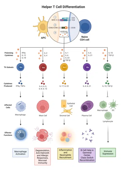
What Cytokines Drive CD4+ TH1 Cell Differentiation?
Understanding the cytokine environment that drives TH1 polarization is essential for designing effective in vitro protocols. Differentiation of naïve CD4+ T cells into TH1 cells is governed by the following signals:
- IL-12 — the primary polarizing cytokine; activates STAT4 and induces IFNγ and T-bet expression
- IFNγ (interferon-gamma) — a key TH1 effector cytokine and positive feedback signal
- IL-2 — promotes T cell proliferation and survival
- Anti-IL-4 blocking antibodies — used to suppress TH2 polarization and maintain TH1 commitment
This cytokine framework mirrors the in vivo signaling environment that drives CD4+ T lymphocyte commitment to the TH1 lineage during infection.
Step-by-Step Protocol: Generating TH1 Cells from Human PBMC
Step 1 — Isolate Naïve CD4+ T Cells
Begin by isolating naïve CD4+ T cells from human PBMC. Magnetic bead-based negative selection is the most common approach and minimizes unwanted activation.
These kits use negative selection to yield an “untouched” population of naïve CD4+ T lymphocytes, ready for downstream activation and polarization. Researchers who want to bypass the isolation process entirely can source freshly isolated human CD4+ T cells directly from Sanguine Bio, pre-qualified from healthy human donors.
Step 2 — Activate CD4+ T Cells Through the TCR Complex
TH1 polarization requires concurrent TCR-mediated activation. Three common approaches for activating naïve CD4+ T cells are:
- Plate-bound antibodies — Coat tissue culture plates with anti-CD3 (clone OKT3) and anti-CD28 antibodies in PBS before seeding cells.
- CD3/CD28 Dynabeads — Use Dynal CD3/CD28 T Cell Expander Dynabeads (Life Technologies) at a 1:1 bead-to-cell ratio.
- Anti-CD3 + autologous monocytes — Coat plates with anti-CD3 alone and supply CD28 co-stimulation via autologous monocytes isolated from PBMC.
Step 3 — Add TH1 Polarizing Cytokines
To drive naïve CD4+ T cells toward the TH1 phenotype, add recombinant human IL-12 — either alone, or at a reduced dose combined with anti-IL-4 blocking antibodies to prevent TH2 skewing. Supplement with recombinant human IL-2 to support T cell expansion.
Refresh media and cytokines every 2–3 days, adjusting frequency based on cell density. As cells expand, the interval between media changes will typically shorten.
Step 4 — Expand or Assay TH1 Cells
Functional TH1 activity, including IFNγ expression, can be detected in as few as 3 days post-activation. For longer-term studies or single-cell clonal assays, cells can be expanded for 2–3 weeks in the presence of IL-2.
How Are CD4+ TH1 Cells Detected by Flow Cytometry?
TH1 cells are most commonly identified by intracellular IFNγ staining after short-term restimulation. The standard detection workflow for CD4+ TH1 cells by flow cytometry includes:
- Restimulate cells for 4–6 hours using plate-bound anti-CD3/CD28, CD3/CD28 Dynabeads, or PMA/ionomycin.
- Add Brefeldin A during restimulation to block cytokine secretion and enable intracellular accumulation.
- Fix and permeabilize cells.
- Stain for cell surface markers (e.g., CD4, CXCR3, CCR5) and intracellular IFNγ and/or T-bet.
This approach allows simultaneous phenotyping and functional characterization of CD4+ T lymphocytes within a single flow cytometry panel.
Non-Polarized Controls: TH0 Conditions
For meaningful experimental comparisons, run parallel cultures under non-polarizing (TH0) conditions. Typically, no exogenous polarizing cytokines are added beyond IL-2. Some protocols also include both anti-IL-12 and anti-IL-4 blocking antibodies, to prevent inadvertent TH1 or TH2 polarization driven by endogenous cytokine production from the T cells themselves.
🧬 Planning a TH1 polarization experiment? Start with high-quality, donor-characterized human CD4+ T cells from Sanguine Bio — available fresh from healthy human donors with documented demographics. Order CD4+ T Cells →
Frequently Asked Questions About CD4+ TH1 Cells
What is the difference between CD4+ T cells and CD8+ T cells?
CD4+ T cells (helper T cells) coordinate immune responses by secreting cytokines and activating other immune cells. CD8+ T cells (cytotoxic T lymphocytes) directly kill infected or cancerous cells. CD4+ T lymphocytes recognize antigens presented on MHC class II molecules, whereas CD8+ T cells recognize antigens on MHC class I.
What are the main subtypes of CD4+ helper T cells?
- TH1 — cell-mediated immunity against intracellular pathogens; secretes IFNγ and IL-2
- TH2 — humoral immunity against extracellular parasites; secretes IL-4, IL-5, IL-13
- TH17 — mucosal defense against bacteria and fungi; secretes IL-17
- Tregs — regulatory suppression of excessive immune responses; expresses FoxP3
- Tfh — support B cell activation in lymphoid follicles
What markers identify TH1 cells?
- T-bet (TBX21) — master transcription factor
- IFNγ — signature cytokine
- CXCR3 — chemokine receptor
- CCR5 — chemokine receptor (also an HIV co-receptor)
- CD4 — lineage marker shared by all helper T cell subsets
How long does it take to generate TH1 cells from PBMC?
Functional TH1 cells can be generated and assayed for IFNγ production in as few as 3 days. For expanded or clonal populations, culture periods of 2–3 weeks with IL-2 supplementation are standard.
Where can I buy human CD4+ T cells for research?
Sanguine Bio offers freshly isolated human CD4+ T cells sourced from healthy, consented donors. Cells are available with donor demographic information, making them suitable for a wide range of immunology research applications.
Summary
Generating CD4+ TH1 helper T cells from human PBMC is a well-established, reproducible protocol. Start with magnetically isolated naïve CD4+ T lymphocytes. TCR activation combined with IL-12 supplementation reliably drives TH1 polarization with high efficiency. When paired with proper non-polarizing TH0 controls and intracellular cytokine staining by flow cytometry, this workflow provides a robust platform for studying CD4+ T cell biology, cytokine responses, and the mechanisms of cell-mediated immunity.
Ready to start your experiment? Sanguine Bio’s freshly isolated human CD4+ T cells are donor-characterized, quality-tested, and ready to use — so you can move from isolation to assay without delay. Shop Human CD4+ T Cells →
Related Reading
- CD4+ T cells in autoimmunity: mechanisms, models, and research considerations
- CD4+ T cells in the tumor microenvironment
- What are CD4+ T cells? A complete guide to CD4 helper T cells
Further Reading
- Zhu J, Yamane H, Paul WE. Differentiation of effector CD4 T cell populations. Annu Rev Immunol. 2010;28:445–89.
- Messi M, et al. Memory and flexibility of cytokine gene expression as separable properties of human TH1 and TH2 lymphocytes. Nat Immunol. 2003;4(1):78–86.
- Volpe E, et al. A critical function for TGF-β, IL-23 and proinflammatory cytokines in driving human TH17 responses. Nat Immunol. 2008;9(6):650–7.