Clinical Significance of CD133 and KLK6 in Malignant Brain Tumors
Why Malignant Brain Tumors Are Hard to Treat
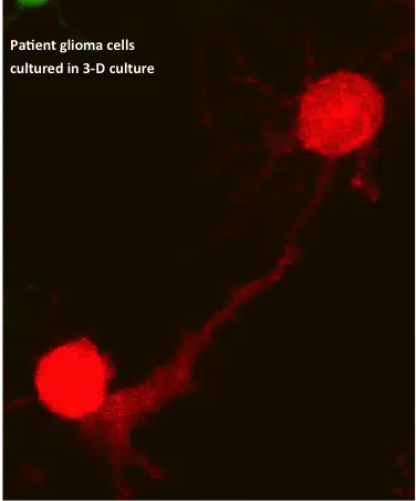
In an earlier article on Glioblastoma Multiforme (GBM), we covered several obstacles that make these lethal brain tumors virtually incurable. Two hallmarks of malignant brain tumors are believed to be the biggest obstacles to killing all tumor cells with current aggressive treatment: glioma cells’ exceptional ability to invade surrounding tissue, and the heterogeneity of GBM — not just between patients, but within a single tumor mass. Both of these characteristics have driven major interest in GBM clinical research.
The World Health Organization (WHO) grades CNS tumors from grade I to grade IV based on how aggressive they are (GBM is WHO grade IV astrocytoma). Most neoplasms spread locally in a limited way and metastasize through the vasculature or lymphatic system. Glioma is different: single glioma cells travel several centimeters through nearby brain tissue but almost never form systemic metastasis. This means WHO tumor grade tracks with proliferation rate rather than tumor invasion — pointing to several independent genetic events driving glioma progression, consistent with the high heterogeneity seen in GBM.
To develop a therapy that finds and destroys malignant glioma cells, researchers need a marker expressed exclusively on the surface of these cells. Because GBM is so heterogeneous, and varies so much between patient brain tumors, no such marker has been identified yet.
Cancer Stem Cells in GBM
Strong evidence points to a subpopulation of malignant cells in GBM with stem-cell-like characteristics: multipotency, and the ability to self-renew and invade. These tumor-initiating cells are called cancer stem cells (CSCs), and they’re believed to drive tumor recurrence in GBM patients. As a result, researchers are moving away from searching for a single mutation or marker. It’s also becoming increasingly important to study primary GBM patient samples directly, since glioma cell lines don’t capture the cellular and molecular makeup of primary GBM.
CD133 as a Tumor Marker
CD133 (also known as Prominin-1 or AC133) is a penta-spanning membrane protein with two heavily glycosylated extracellular loops. It’s recognized as a stem cell marker in certain normal and cancerous tissues, accumulating near the Golgi and ER as well as on the cell surface. Many earlier studies reported that a CD133+ glioma CSC subpopulation drives tumor formation and rapid proliferation in GBM. But later, conflicting findings showed that CD133-negative glioma cells can also self-renew and form tumors in xeno-transplantation assays.
Recent findings from Brescia’s group confirm that cell-surface CD133 expression marks self-renewing, tumor-initiating GBM cells — but it isn’t essential for stem cell properties in every GBM case. Membrane-bound CD133 was only detectable in a fraction of patient samples (neurospheres and freshly dissociated tumors). Yet CD133 mRNA and intracellular CD133 protein were found at high levels in nearly all the neurosphere samples tested.
Through clonal analysis, the researchers traced the behavior of single glioma cells and their progeny. Every clone from a single CD133-negative cell contained a mix of both CD133-negative and CD133-positive cells. They also found that cell-surface CD133 shifts back and forth between the cytoplasm and the plasma membrane — likely controlled by cues from the tumor’s microenvironment.
These findings move GBM research a step closer to understanding how malignant gliomas progress. But the existence of a cytoplasmic CD133 reservoir, and its recycling to the plasma membrane and back, limits how useful CD133 can be as a glioblastoma-targeting marker.
KLK6 and Glioma Survival
In a separate study published the same week in Neuro-Oncology, researchers at Mayo Clinic found a significant link between malignant gliomas and the Kallikrein 6 (KLK6) enzyme. KLK6 belongs to the kallikrein family of secreted serine proteases and is known to rise in areas of CNS inflammation, suggesting it’s regulated along with T-cell activation. Notably, serum from Multiple Sclerosis (MS) patients also shows elevated KLK6 levels. The study found that as KLK6 expression rises, post-surgery survival time for GBM patients falls — with the highest KLK6 levels found in the most severe GBM cases. These results came from looking at 60 samples of grade IV astrocytomas (classified as GBM) and less aggressive grade III astrocytomas.
Scarisbrick’s group also showed that KLK6 may help malignant glioma cells survive and resist apoptosis-inducing treatments like radiotherapy (RT) and temozolomide (TMZ). This pro-survival effect of KLK6 could point to a new GBM treatment strategy — though the supporting experiments were done in the U251 glioma cell line. If future studies confirm that KLK6 also promotes survival in primary patient tumor cells, a therapy targeting KLK6 could become a promising addition to GBM treatment after surgical resection and before chemo- and radiotherapy.
Further Reading
- CD133 is essential for glioblastoma stem cell maintenance.
- CD133 as a marker for regulation and potential for targete therapies in glioblastoma multiforme.
- Clinical significance and novel mechanism of action of kallikrein 6 in glioblastoma.
- Functional role of kallikrein 6 in regulating immune cell survival.