ISSCR 2013 Meeting Updates: Can Alzheimer’s Disease be modeled in a dish?

Alzheimer’s Disease: Two Pathologies, One Open Question
Human pluripotent stem cells (hPSCs) can differentiate into every cell type in the body, making them powerful models for studying the pathological mechanisms behind human disease. At the International Society for Stem Cell Research (ISSCR) 11th Annual Meeting, stem cell experts presented the current human stem cell models for Alzheimer’s disease and discussed where the field is headed next.
Alzheimer’s disease (AD) is the most common neurodegenerative dementia, affecting roughly 30 million people worldwide. AD occurs in two main forms: early-onset, familial AD (FAD), and late-onset, sporadic AD (SAD). 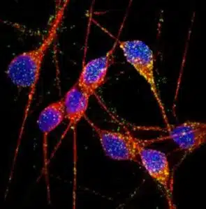 Both forms are marked by extensive neuronal loss and the aggregation of two proteins in the brain: amyloid β peptide (Aβ) and tau. Aβ peptide comes from the amyloid precursor protein (APP), cleaved by two proteases, β-secretase and γ-secretase. According to the amyloid cascade hypothesis, elevated Aβ levels are necessary and sufficient to trigger disease1.
Tau is made in neurons, where it normally binds tubulin and stabilizes microtubules. In AD, though, tau becomes hyper-phosphorylated. This causes it to detach from microtubules, aggregate, and form neurofibrillary tangles (NFTs). Amyloid plaques and NFTs are the two pathological hallmarks of AD, but exactly how they relate to each other — and how they drive the onset and progression of AD — is still under investigation. By the time a patient shows symptoms of mild dementia, significant neuronal loss and substantial plaque and tangle buildup have already occurred. One major limitation in AD research has been the lack of live, patient-specific neurons for studying disease progression.
Modeling Familial and Sporadic AD with iPSCs
Advances in reprogramming technology now let scientists generate induced pluripotent stem cells (iPSCs), creating live, patient-specific models to study disease phenotypes in a dish. At the ISSCR meeting, Larry Goldstein presented his lab’s work using hiPSC models to study AD. His team generated iPSCs from two patients with FAD caused by a duplication of the APP gene, two patients with SAD, and two control individuals. They then generated neurons from these iPSC lines through directed differentiation and fluorescence-activated cell sorting (FACS) purification2.
Neurons from one SAD patient and two FAD patients showed significantly higher levels of secreted Aβ and phosphorylated tau (p-tau)3. To test whether APP processing was linked to elevated p-tau levels, the team treated iPSC-derived neurons with γ-secretase and β-secretase inhibitors. Blocking β-secretase significantly reduced both Aβ and p-tau levels. Blocking the γ-secretase inhibitor only reduced Aβ levels, not p-tau. This suggests that APP processing byproducts other than Aβ may contribute to elevated p-tau — a finding that points to a potential weakness in the amyloid cascade hypothesis.
An Alternative Hypothesis: Intracellular Aβ Oligomers
Other research groups have proposed alternative explanations for AD pathogenesis. Haruhisa Inoue presented his group’s work using human iPSC models to study how intracellular Aβ oligomers contribute to AD. They generated iPSCs from one FAD patient carrying the APP-E693Δ mutation, two SAD patients, and three control individuals, then derived cortical neurons using small-molecule inhibitors of bone morphogenic protein (BMP) and activin/nodal signaling, following a previously described method4.
Aβ oligomers built up in neurons from the FAD patient and one SAD patient, but not in control neurons5. Specifically, the oligomers accumulated in the endoplasmic reticulum (ER), triggering ER and oxidative stress in the neurons. Treatment with docosahexaenoic acid (DHA) eased these stress responses. The drug has failed in some previous AD clinical trials, but Inoue’s work suggests DHA might still help a subset of patients.
What This Means for AD Research
Goldstein and Inoue both presented convincing evidence that human iPSC models can be used to study early AD pathogenesis and patient-specific drug responses. Disease phenotypes can show up in iPSC models even though it typically takes decades for symptoms to appear in patients. That said, only one of the two SAD patients in Goldstein’s study generated a disease phenotype. This highlights the need for future iPSC studies to include larger numbers of patients, to account for the heterogeneity seen in AD pathogenesis.
References
1 Hardy, J. & Selkoe, D. J. The amyloid hypothesis of Alzheimer’s disease: progress and problems on the road to therapeutics. Science 297, 353-356, doi:10.1126/science.1072994 (2002).
2 Yuan, S. H. et al. Cell-surface marker signatures for the isolation of neural stem cells, glia and neurons derived from human pluripotent stem cells. PLoS One 6, e17540, doi:10.1371/journal.pone.0017540 (2011).
3 Israel, M. A. et al. Probing sporadic and familial Alzheimer’s disease using induced pluripotent stem cells. Nature 482, 216-220, doi:10.1038/nature10821 (2012).
4 Morizane, A., Doi, D., Kikuchi, T., Nishimura, K. & Takahashi, J. Small-molecule inhibitors of bone morphogenic protein and activin/nodal signals promote highly efficient neural induction from human pluripotent stem cells. J Neurosci Res 89, 117-126, doi:10.1002/jnr.22547 (2011).
5 Kondo, T. et al. Modeling Alzheimer’s disease with iPSCs reveals stress phenotypes associated with intracellular Abeta and differential drug responsiveness. Cell Stem Cell 12, 487-496, doi:10.1016/j.stem.2013.01.009 (2013).