
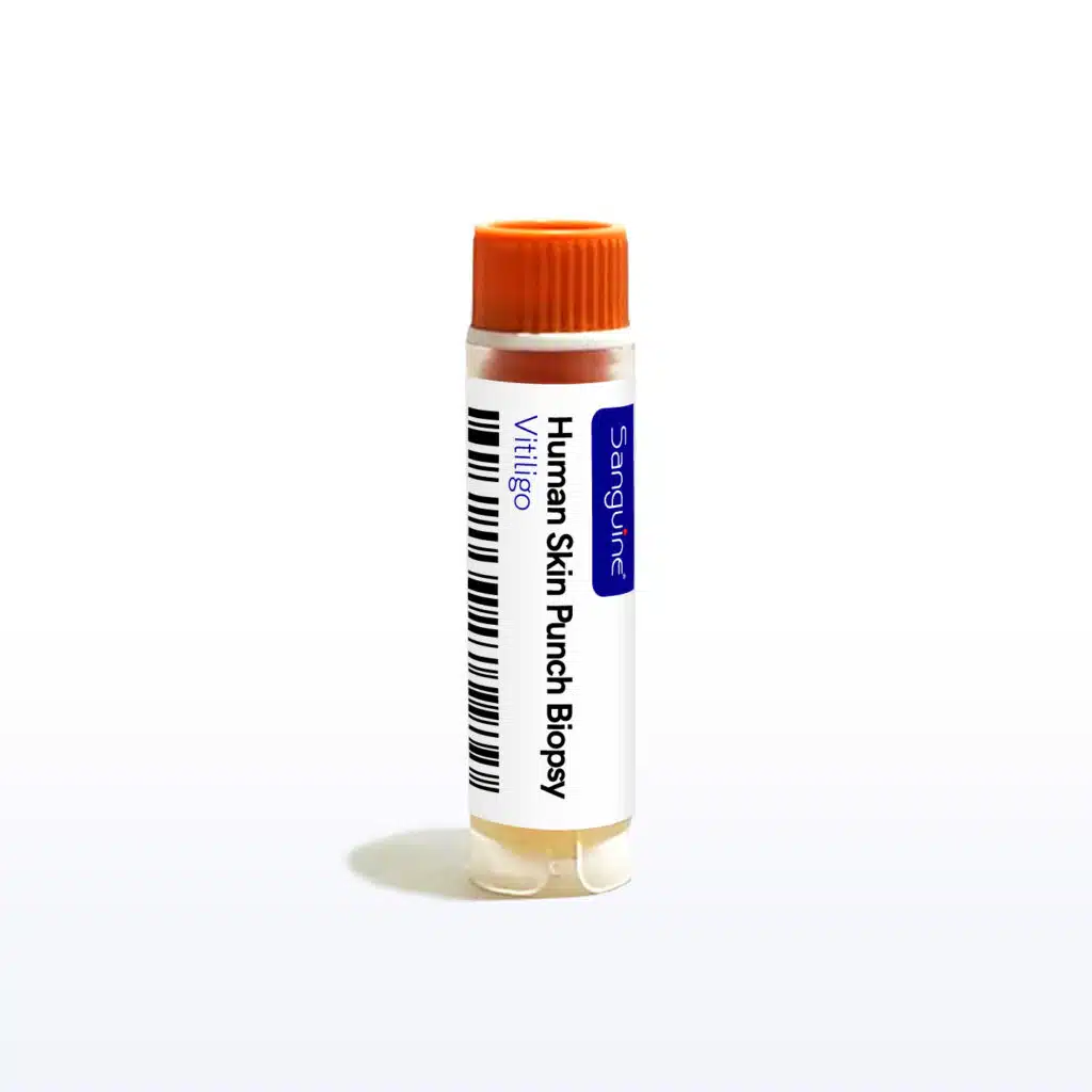
Vitiligo Skin Punch Biopsy
About:Vitiligo Skin Punch Biopsy samples are collected from consented donors with clinically confirmed vitiligo, an autoimmune disease of melanocyte destruction, with lesional, perilesional, and non-lesional sites available. Suitable for quantifying melanocyte loss, imaging CD8+ T cell infiltrates at the lesional margin, profiling IFN-gamma-driven chemokines and JAK-STAT activation, and characterising resident memory T cells. Matched healthy controls available.
About Vitiligo Skin Punch Biopsy
Vitiligo Skin Punch Biopsy for Autoimmune Depigmentation Research
Vitiligo Skin Punch Biopsy samples are collected from IRB-consented donors with a clinically confirmed diagnosis of vitiligo. Biopsies are taken by licensed clinicians under aseptic conditions and, where frozen material is requested, are frozen within 4 hours of collection. Lesional depigmented, perilesional, and clinically normal non-lesional sites can be requested. These specimens support histological, immunological, and molecular research into melanocyte loss and the immune response that causes it.
Why Skin Tissue for Vitiligo Research
Vitiligo is an autoimmune disease in which epidermal melanocytes are destroyed by autoreactive cytotoxic T cells recognising melanocyte differentiation antigens. Both the target cell and the effector cells reside in the epidermis, so skin tissue is the only specimen in which the disease process itself can be observed.
- Melanocyte loss is defined histologically: lesional epidermis shows absence of melanocytes on immunostaining for markers such as Melan-A, SOX10, or MITF, and absence of melanin pigment. This is the primary endpoint for repigmentation research.
- CD8+ cytotoxic T cells accumulate at the advancing lesional margin, which is why perilesional biopsies are taken separately from stable depigmented centres — the two sites report on different phases of the disease.
- The IFN-gamma–CXCL9/CXCL10–CXCR3 axis recruits autoreactive T cells into the epidermis, and these chemokines are measurable in lesional tissue. Signalling downstream of IFN-gamma runs through JAK-STAT, which is the mechanistic basis for JAK inhibitor therapy in vitiligo.
- Autoreactive resident memory CD8+ T cells persist in lesional and in clinically normal-appearing skin and are implicated in relapse after treatment withdrawal; these cells are recoverable only from tissue, not from blood.
- Melanocyte-intrinsic oxidative and cellular stress responses contribute to susceptibility, and these are assessed in the melanocyte and keratinocyte compartments in situ.
- Paired lesional and non-lesional biopsies from the same donor provide an internally controlled comparison, separating disease-driven change from inter-individual and skin-type variation.
Donor Stratification Available
- Subtype: nonsegmental (generalised or acrofacial), segmental, or universal vitiligo
- Disease activity: stable versus actively spreading, including confetti-like depigmentation, trichrome lesions, or Koebner phenomenon where documented
- Biopsy site: lesional depigmented centre, perilesional advancing margin, or clinically normal non-lesional skin
- Extent of involvement and body surface area or VASI score where recorded
- Treatment status: topical corticosteroid, topical calcineurin inhibitor, JAK inhibitor, narrowband UVB phototherapy, or treatment-naive
- Concurrent autoimmune conditions, notably autoimmune thyroid disease, and Fitzpatrick skin type
- Matched healthy controls available
Product Features
- Research Use Only (RUO), fresh or frozen skin punch biopsies
- Samples are frozen (if requested) within 4 hours of collection
- Clinically confirmed vitiligo donors
- Collected by licensed clinicians under aseptic conditions
- Lesional, perilesional, and non-lesional sites available from the same donor on request
- Processed and shipped same-day where applicable
- IRB-approved protocols and electronic informed consent
De-identified Donor Data
- Verified diagnosis of vitiligo and subtype where documented
- Donor demographics: age, sex assigned at birth, race/ethnicity
- Self-reported medication history and lesion site
- Disease activity, extent, and clinical history available upon request
Applications
- Immunohistochemical quantification of melanocyte density and pigment loss
- Multiplex imaging of CD8+ T cell infiltrates at the lesional margin
- Characterisation of resident memory T cell populations in lesional and non-lesional skin
- Measurement of IFN-gamma-inducible chemokines and JAK-STAT pathway activation in situ
- Spatial and single-cell transcriptomic profiling of epidermal and dermal compartments
- Target validation for JAK inhibitor, IL-15, and CXCR3-directed therapeutic programmes
- Melanocyte stress response and oxidative damage studies
- Repigmentation and melanocyte regeneration research, including follicular melanocyte reservoirs
Other Vitiligo Specimen Types
Compliance and Quality Assurance
- IRB-approved collections and SOP-driven processing
- 21 CFR Part 11-compliant electronic informed consent
- HIPAA-compliant donor data management
Ordering & Customization
Vitiligo Skin Punch Biopsy samples can be shipped ambient, refrigerated, or on dry ice. For pricing, availability, orders outside the United States, or regulatory documentation, please contact learnmore@sanguinebio.com. To browse other disease states and biopsy formats, visit our human skin punch biopsy page.
Applications
- Immunology and translational research
- Biomarker discovery and validation
- Drug screening and assay development
- Vaccine and infectious disease research
Product Features
- Research Use Only (RUO), Skin Punch Biopsy
- Sourced from screened, consented donors
- Processed under validated SOPs
- QC tested for purity, viability, and sterility
- Processed within 24 hours of collection
- IRB-approved protocols with electronic informed consent
Donor Metadata
- Verified diagnosis and clinical history
- Demographic data: age, sex assigned at birth, race/ethnicity
- Medication and treatment background when available
Compliance and Quality Assurance
- IRB-approved collections and standardized procedures
- 21 CFR Part 11-compliant e-consent system
- HIPAA-compliant data management
Ordering & Customization
Human Skin Punch Biopsy are available in customizable aliquot sizes. For international orders or documentation needs, please contact learnmore@sanguinebio.com to confirm requirements and availability. We also work with multiple ordering platforms and distributors, learn more here.
Where can I find your complete catalog?
You can find our full product catalog here.
How long can samples be stored?
Storage depends on sample type but often cryopreserved samples stored in liquid nitrogen can be stored for years.
What customization options are available?
Extensive customization is available. To discuss your project request a quote or email us at learnmore@sanguinebio.com.
Do you offer prospective collections?
Sanguine offers flexible prospective collection services tailored to fit your research. For more information and to request a quote, please see: our prospective biospecimen collection services page.
What is the turnaround time for custom collections?
Timelines depend on condition, sample type, and I/E criteria but we often start collection within 2 weeks of signed agreement.
Do you have samples in stock?
YES - we have in-stock inventory. You can also email us at learnmore@sanguinebio.com as we continually get new samples in our inventory.
What does unique donor mean?
Each sample from a different individual - Unique donor means each sample comes from a different person, ensuring biological diversity in your study.
Are samples collected under IRB-approved protocols?
YES - "IRB-approved collection protocols" and IRB approval documentation available upon request. Sanguine utilizes two internationally-recognized IRBs (Advarra and WCG IRB) for review and approval.
Frequently Asked Questions
Ask a Question
Pair with a Healthy Control
Match your Vitiligo Skin Punch Biopsy with normal donor samples for comparison
Healthy Skin Punch Biopsy
View InventoryNeed a custom Skin Punch Biopsy cohort?
Our scientific team can scope prospective collections with donor-specific I/E criteria, typically starting within 2 weeks of agreement.