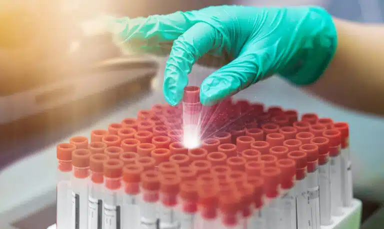
Metabolomics and Small Molecule Profiling: Sample Collection Considerations
Metabolomics is the full-scale analysis of small molecules (<1,500 Da) — amino acids, lipids, carbohydrates, organic acids, nucleotides, and xenobiotic metabolites. It gives systems-level insight into cellular metabolism, disease biology, and treatment response. Genomics shows a static blueprint, and transcriptomics captures intermediate states. Metabolomics reflects the actual functional outcome, combining genetic, transcriptional, and post-translational regulation with outside influences like diet, microbiome, and drug exposure.[1]
This closeness to phenotype makes metabolomics powerful for biomarker discovery, mechanistic work, and precision medicine across nearly every disease area. But that same closeness to phenotype makes it uniquely vulnerable to handling artifacts. Metabolite levels turn over fast and can shift within seconds to minutes of collection.
The human metabolome includes an estimated 100,000+ chemical entities, from simple molecules like glucose and lactate to complex, many-carbon lipids. Mass spectrometry-based metabolomics routinely measures 500–2,000 metabolites per sample across central carbon metabolism, amino acid pathways, lipid species, nucleotide pools, and specialized metabolites.[2]
Different metabolite classes have very different stability. Some stay stable for days at room temperature; others degrade within minutes unless specimens are processed and frozen right away. That variation calls for tight standardization of collection, processing, and storage, so the measured profile reflects true biology rather than handling artifacts.
At Sanguine, our metabolomics expertise covers protocol optimization across specimen types and analytical platforms. Human Plasma collected in EDTA tubes with immediate cold processing preserves most metabolite classes. Human Serum offers an alternative matrix for applications where clotting activates metabolically relevant pathways. Specialized protocols use metabolite stabilizers, rapid quenching, and temperature control to limit metabolic activity outside the body.[3] Our distributed processing network across the United States keeps collection-to-freezer timelines under 4 hours for labile metabolites. Each sample also comes with detailed genomic annotation from study design to receipt of samples.
Pre-Analytical Variables in Metabolomics
Collection Timing and Patient Preparation
Circulating metabolite levels shift with food intake, activity, circadian rhythm, and stress. Without standardized collection conditions, that creates a lot of pre-analytical variability.
Postprandial changes are especially dramatic. Glucose, insulin, amino acids, and lipids can all rise 50–500% within 1–4 hours of a meal.[4] These are normal responses to eating, not signs of disease, but they confound studies performed at inconsistent postprandial states. Fasting collection (typically an 8–12 hour overnight fast) removes these acute swings and captures a basal metabolic state.
Circadian rhythms affect many metabolites, including cortisol, melatonin metabolites, and certain amino acids, with 2–3 fold morning-to-evening swings for some.[5] Standardizing collection to one time of day (typically 8–10 AM) minimizes this effect. Studies specifically investigating circadian metabolism may instead sample at multiple timepoints over 24 hours under carefully controlled conditions.
Medications alter metabolite profiles two ways: directly, through drug-metabolite contributions, and indirectly, through their effect on the body’s own metabolism.[6] Ideally, subjects pause medications for a washout period before baseline profiling. That’s often impractical or unethical for patients on continuous therapy, though. Thorough medication documentation — drug names, dosages, and timing relative to collection — lets researchers correct for this or run subgroup analyses later.
Anticoagulant Selection
The choice of plasma anticoagulant strongly shapes metabolomic profiles. It can interfere with analytical platforms, contribute its own metabolites, and affect residual cellular activity during processing differently.
EDTA plasma is the most widely used matrix for metabolomics, with strong stability and minimal interference for most mass spectrometry platforms.[7] One caveat: EDTA binds divalent cations, including calcium and magnesium, which can affect certain enzymatic reactions and create artificial shifts in specific metabolites.
Anticoagulant Comparison for Metabolomics:
- EDTA Plasma: Preferred for most applications; minimal interference, good stability; our standard Human Plasma collection.
- Heparin Plasma: Alternative matrix; preserves calcium-dependent pathways, but shows batch-to-batch variability and may interfere with some LC-MS methods.
- Citrate Plasma: 10% dilution from anticoagulant; good for coagulation-related metabolites; less common for general metabolomics.
- Serum: Clotting activates platelets and coagulation cascades, releasing metabolites; useful for specific applications but introduces variability.
- Fluoride/Oxalate: Glycolysis inhibitors that preserve glucose; not recommended for comprehensive metabolomics due to enzyme inhibition artifacts.
Serum metabolomics captures metabolites released during clotting, including platelet-derived factors and coagulation intermediates.[8] Serum works well for studying those processes specifically. But clotting adds temporal variability — typically 30–60 minutes of clot formation, during which continued cellular activity keeps changing the profile. Human Serum applications include platelet metabolism, serotonin pathway analysis, and studies where clotting physiology matters.
Processing Speed and Temperature Control
Metabolite stability varies enormously by class. Energy metabolites like ATP have half-lives measured in minutes, while structural lipids stay stable for hours at room temperature. Fast processing, minimizing the gap between blood draw and plasma separation, is critical for preserving labile pools.[9] Our Human Plasma is processed within 30–60 minutes of collection, with immediate centrifugation at 4°C to slow enzymatic activity and prevent glycolysis and oxidation.
Blood cells left in contact with plasma keep metabolizing. They consume glucose, produce lactate, consume oxygen, and release internal metabolites as membranes become more permeable.[10] These processes create artificial shifts that misrepresent true circulating levels. Rapid separation removes more than 99.9% of cells and stops these processes. Even cell-free plasma retains some enzymatic activity, so freezing right away at −80°C stops most remaining degradation, though some change happens during the freeze-thaw itself.
Metabolite Stability and Storage Considerations
Temperature affects metabolite stability at every step, from collection through long-term storage. Some metabolites tolerate room temperature for hours; others need immediate ice-water immersion.[11] Standard protocols typically use room-temperature transport followed by immediate cold processing, though specific analytes need alternatives. Ascorbic acid (vitamin C) oxidizes fast and needs metaphosphoric acid added at collection. Polyunsaturated fatty acids, which oxidize easily, benefit from antioxidant addition (BHT, EDTA).
Metabolite Class-Specific Stability Considerations:
- Energy metabolites (ATP, ADP, AMP): Highly labile, degrade within minutes, require immediate freezing
- Glycolytic intermediates: Unstable; glycolysis continues outside the body; fluoride/oxalate tubes or immediate processing essential
- Amino acids: Generally stable; can tolerate 1–2 hours at room temperature
- Lipids: Most classes stable, though polyunsaturated species oxidize slowly; antioxidants recommended
- Acylcarnitines: Stable in plasma for several hours
- Organic acids: Variable stability; some stable, others degrade within hours
- Methylated metabolites: Generally stable
- Nucleotides: Relatively unstable; rapid processing recommended
- Vitamins: Variable (vitamin C highly labile, vitamin D stable)
Freeze-thaw cycles progressively degrade many metabolites — through enzymatic activity during thaw, protein precipitation that exposes metabolites to matrix effects, and physical stress that causes fragmentation.[12] Single-use aliquots eliminate this variability. Small aliquots (50–200 µL), sized for specific assays, avoid thawing an entire sample per analysis. Our Human Plasma is aliquoted into multiple small portions, so longitudinal collections can be accessed repeatedly without freeze-thaw degradation.
Long-term storage stability also varies by class. Some metabolites stay stable for decades at −80°C, while others show detectable degradation within months. Periodic stability checks on archived specimens confirm whether conclusions still hold years after banking.[13] Quality control samples stored alongside research specimens allow monitoring through periodic re-analysis. Any significant concentration change then prompts a closer look at storage conditions or analytical drift.
Specimen Types for Metabolomics Applications
Plasma vs Serum Selection
Plasma is the preferred matrix for most metabolomics work: it processes faster, avoids clotting-related artifacts, and stays stable longer for many classes. Plasma’s roughly 15% higher yield compared to serum (serum excludes clotted volume) can matter when sample volume is limited.[14] EDTA plasma performs well across LC-MS and GC-MS platforms, with minimal ion suppression or chromatographic interference.
Serum works well for specific applications where clotting releases biologically relevant metabolites, or where established clinical assays already use serum. Lipidomics sometimes uses serum, since many lipid species tolerate clotting and historical lipid reference ranges were set in serum.[15] Serotonin studies may also prefer serum, since platelets release serotonin during clotting, which reflects whole-blood content rather than the low levels found in platelet-poor plasma.
Urine Metabolomics
Urine complements blood: it’s enriched in water-soluble metabolites the kidneys excrete, and depleted in lipophilic species that stay in circulation. It’s less invasive than blood, allows longitudinal sampling without medical staff, and captures kidney-filtered and secreted metabolites that reflect organ-specific processes.[16] Concentrations vary widely with hydration, so creatinine normalization corrects for dilution across samples and timepoints.
Urine Collection Considerations:
- First morning void preferred (overnight accumulation, consistent timing)
- Mid-stream clean catch minimizing bacterial contamination
- Immediate refrigeration or freezing after collection
- Centrifuge to remove particulates and cells before analysis
- Aliquot into small volumes for single-use
- Measure creatinine for normalization
- Record collection timing and any medications/supplements
- Avoid protease inhibitors (may interfere with downstream analyses)
- Store at −80°C for long-term banking
Tissue and Cellular Metabolomics
Tissue biopsies and isolated cells allow metabolomic analysis of specific cell types or organs, revealing compartment-specific metabolism that’s invisible in bulk plasma. But ischemia after excision quickly depletes energy metabolites and triggers stress responses if processing is delayed.[17] Immediate snap-freezing in liquid nitrogen stops metabolism within seconds. Human PBMCs metabolomics reveals immune cell metabolic changes in disease, and rapid processing and quenching are essential for accurate energy-metabolite measurement.
Analytical Platform Considerations
Targeted vs Untargeted Metabolomics
Targeted metabolomics measures predefined panels using optimized methods with internal standards for absolute concentrations. It offers strong quantitative accuracy, lower detection limits, and excellent reproducibility, which makes it ideal for biomarker validation and clinical applications.[18] The trade-off: it misses unexpected or novel metabolites outside the panel. Common targeted panels measure amino acids (20–40 species), acylcarnitines (20–60), organic acids (30–50), and biogenic amines including neurotransmitter metabolites.
Untargeted metabolomics uses high-resolution mass spectrometry to capture thousands of features without assuming which metabolites are present. This discovery-oriented approach finds unexpected changes, novel biomarkers, and pathway shifts that targeted methods would miss.[19] The trade-offs: identification requires database matching and often remains tentative without confirmatory standards, and quantification is only semi-quantitative without internal standards. Untargeted workflows benefit from large sample sets for statistical power.
Sample Preparation Methods
Protein precipitation is the most common preparation method. It uses organic solvents (methanol, acetonitrile) to precipitate proteins while pulling small molecules into the liquid above. It’s fast, scalable, and works with most LC-MS platforms.[20] The downside is that it co-extracts lipids, which can suppress ion signal in electrospray ionization. Solid-phase extraction (SPE) selectively enriches specific metabolite classes, improving sensitivity for targeted work at the cost of added complexity and sample volume.
Derivatization converts non-volatile metabolites into volatile derivatives for GC-MS, which is especially valuable for organic acids, amino acids, and sugars. Common agents include trimethylsilyl and methoxime reagents.[21] It adds processing steps and potential artifacts, but covers parts of the metabolome that complement LC-MS.
Quality Control for Metabolomics Studies
Solid QC matters given how sensitive metabolomics is to pre-analytical variables and analytical drift. Pooled QC samples, made by combining small aliquots from all study specimens, give matrix-matched controls that span the full concentration range of the study population.[22] Analyzed repeatedly throughout a batch, these pooled QCs catch sensitivity drift, retention-time shifts, and other technical variation that needs correcting before biological interpretation.
Essential QC Elements:
- Pooled biological QC samples analyzed every 8–12 injections
- Commercial reference standards for retention time and mass calibration
- Internal standards added to all samples correcting for extraction efficiency
- Blank injections detecting carryover or contamination
- Coefficient of variation monitoring (<20% for most metabolites)
- Principal component analysis identifying outlier samples
- Batch effect assessment and correction when multi-batch analysis required
- Normalization methods accounting for total signal intensity variations
- Missing value imputation strategies for below-detection-limit metabolites
External quality assessment programs allow cross-laboratory comparison, showing whether a lab’s results match field-wide consensus. Taking part in proficiency testing documents analytical competence and surfaces systematic biases that need troubleshooting.[23]
Metabolite Databases and Pathway Analysis
Identifying metabolites from mass spectral data requires databases that catalog neutral masses, fragmentation patterns, retention times, and collision cross-sections. HMDB (Human Metabolome Database), METLIN, and LipidMaps are gold-standard resources that link analytical signatures to chemical identities.[24] Many detected features still remain unidentified — unknown metabolites, drug metabolites, or environmental exposures not yet characterized.
Pathway analysis maps identified metabolites onto biochemical pathways, revealing coordinated regulation that’s invisible from looking at individual changes alone. KEGG, Reactome, and MetaboAnalyst enable statistical assessment of pathway-level changes.[25] Combining this with transcriptomics and proteomics gives systems-level insight across molecular layers.
Disease-Specific Metabolomic Approaches
Cancer Metabolism
Cancer cells show major metabolic reprogramming: the Warburg effect (aerobic glycolysis), glutamine addiction, and altered lipid metabolism that support rapid growth. Plasma metabolomics from cancer patients reveals these systematic changes as circulating signatures. Elevated lactate, altered amino acid ratios, and disrupted lipids distinguish cancer from healthy controls.[26] Our Human Plasma from various cancer types shows disease-specific fingerprints that correlate with tumor burden, stage, and treatment response.
Inborn Errors of Metabolism
Genetic defects in metabolic enzymes cause substrate buildup and product shortages, which newborn screening and diagnostic metabolomics can detect. Tandem mass spectrometry of dried blood spots or Human Plasma identifies dozens of inborn errors, including amino acidopathies, organic acidurias, and fatty acid oxidation disorders.[27] Carrier screening and prenatal testing use similar methods.
Cardiovascular Disease
Metabolomic studies of cardiovascular disease have identified circulating biomarkers that predict events years before symptoms appear. Trimethylamine N-oxide (TMAO), made from gut microbiota metabolism of dietary choline, is linked to atherosclerosis risk. Branched-chain amino acids correlate with insulin resistance and diabetes risk.[28] Comprehensive lipidomics from Human Plasma or Human Serum reveals hundreds of lipid species with potential prognostic value.
Multi-Omic Integration
Modern systems biology combines metabolomics with genomics, transcriptomics, and proteomics to characterize biological states across molecular layers. Network analysis identifies correlations between metabolites, transcripts, and proteins, revealing regulatory relationships and feedback loops.[29] Machine learning models trained on multi-omic data can predict phenotypes, classify disease subtypes, and forecast treatment responses more accurately than single-modality approaches. Multi-omic studies need careful specimen planning so every molecular layer stays accessible. Our Human Plasma, Human Serum, and Human PBMCs support comprehensive multi-omic characterization from a single collection event.
Check Our Inventory
Advancing metabolomics research with optimally processed biospecimens?
Sanguine offers Human Plasma and Human Serum collected and processed using metabolomics-optimized protocols. Rapid processing, controlled temperature, and comprehensive genomic annotation support cutting-edge metabolomic investigations from study design to receipt of samples.
Ethical Sourcing and Regulatory Compliance
All Sanguine biospecimens are collected under IRB-approved protocols with full informed consent from every donor. Our HIPAA-compliant data management systems protect donor privacy while giving researchers access to detailed genomic annotation for their studies. We maintain ISO 9001:2015 and ISO 13485:2016 certifications, reflecting our commitment to quality management across all operations.
Donor compensation follows ethical guidelines set by professional societies, ensuring voluntary participation without coercion. Geographic diversity in our collection network across the United States supports health equity in research while giving access to populations often left out of biomedical studies. Every specimen is designated Research Use Only (RUO) with clear documentation of its intended application scope.
References
- Johnson CH, Ivanisevic J, Siuzdak G. Metabolomics: beyond biomarkers and towards mechanisms. Nat Rev Mol Cell Biol. 2016;17(7):451-459. doi:10.1038/nrm.2016.25
- Wishart DS. Emerging applications of metabolomics in drug discovery and precision medicine. Nat Rev Drug Discov. 2016;15(7):473-484. doi:10.1038/nrd.2016.32
- Kamlage B, Maldonado SG, Bethan B, et al. Quality markers addressing preanalytical variations of blood and plasma processing identified by broad and targeted metabolite profiling. Clin Chem. 2014;60(2):399-412. doi:10.1373/clinchem.2013.211979
- O’Sullivan A, Gibney MJ, Brennan L. Dietary intake patterns are reflected in metabolomic profiles. Am J Clin Nutr. 2011;93(2):314-321. doi:10.3945/ajcn.110.000950
- Dallmann R, Viola AU, Tarokh L, et al. The human circadian metabolome. Proc Natl Acad Sci USA. 2012;109(7):2625-2629. doi:10.1073/pnas.1114410109
- Rinschen MM, Ivanisevic J, Giera M, Siuzdak G. Identification of bioactive metabolites using activity metabolomics. Nat Rev Mol Cell Biol. 2019;20(6):353-367. doi:10.1038/s41580-019-0108-4
- Breier M, Wahl S, Prehn C, et al. Targeted metabolomics identifies reliable and stable metabolites in human serum and plasma samples. PLoS One. 2014;9(2):e89728. doi:10.1371/journal.pone.0089728
- Yin P, Lehmann R, Xu G. Effects of pre-analytical processes on blood samples used in metabolomics studies. Anal Bioanal Chem. 2015;407(17):4879-4892. doi:10.1007/s00216-015-8565-x
- Wedge DC, Allwood JW, Dunn W, et al. Is serum or plasma more appropriate for intersubject comparisons in metabolomic studies? Anal Chem. 2011;83(17):6689-6697. doi:10.1021/ac201221h
- Barri T, Dragsted LO. UPLC-ESI-QTOF/MS and multivariate data analysis for blood plasma and serum metabolomics: effect of experimental artefacts and anticoagulant. Anal Chim Acta. 2013;768:118-128. doi:10.1016/j.aca.2013.01.015
- Pinto J, Domingues MR, Galhano E, et al. Human plasma stability during handling and storage: impact on NMR metabolomics. Analyst. 2014;139(5):1168-1177. doi:10.1039/c3an02188b
- Fliniaux O, Gaillard G, Lion A, et al. Influence of common preanalytical variations on the metabolic profile of serum samples in biobanks. J Biomol NMR. 2011;51(4):457-465. doi:10.1007/s10858-011-9574-5
- Anton G, Wilson R, Yu ZH, et al. Pre-analytical sample quality: metabolite ratios as an intrinsic marker for prolonged room temperature exposure of serum samples. PLoS One. 2015;10(3):e0121495. doi:10.1371/journal.pone.0121495
- Hirayama A, Sugimoto M, Suzuki A, et al. Effects of processing and storage conditions on charged metabolomic profiles in blood. Electrophoresis. 2015;36(18):2148-2155. doi:10.1002/elps.201400600
- Siskos AP, Jain P, Römisch-Margl W, et al. Interlaboratory reproducibility of a targeted metabolomics platform for analysis of human serum and plasma. Anal Chem. 2017;89(1):656-665. doi:10.1021/acs.analchem.6b02930
- Bouatra S, Aziat F, Mandal R, et al. The human urine metabolome. PLoS One. 2013;8(9):e73076. doi:10.1371/journal.pone.0073076
- Lin S, Hoffmann TJ, Storb R, et al. An optimized method for detection of primary and secondary metabolites in diverse biological matrices using gas chromatography time-of-flight mass spectrometry. Methods Mol Biol. 2014;1198:79-103. doi:10.1007/978-1-4939-1258-2_6
- Roberts LD, Souza AL, Gerszten RE, Clish CB. Targeted metabolomics. Curr Protoc Mol Biol. 2012;Chapter 30:Unit 30.2.1-24. doi:10.1002/0471142727.mb3002s98
- Patti GJ, Yanes O, Siuzdak G. Innovation: metabolomics: the apogee of the omics trilogy. Nat Rev Mol Cell Biol. 2012;13(4):263-269. doi:10.1038/nrm3314
- Bruce SJ, Tavazzi I, Parisod V, et al. Investigation of human blood plasma sample preparation for performing metabolomics using ultrahigh performance liquid chromatography/mass spectrometry. Anal Chem. 2009;81(9):3285-3296. doi:10.1021/ac8024569
- Fiehn O, Kopka J, Dörmann P, et al. Metabolite profiling for plant functional genomics. Nat Biotechnol. 2000;18(11):1157-1161. doi:10.1038/81137
- Dunn WB, Broadhurst D, Begley P, et al. Procedures for large-scale metabolic profiling of serum and plasma using gas chromatography and liquid chromatography coupled to mass spectrometry. Nat Protoc. 2011;6(7):1060-1083. doi:10.1038/nprot.2011.335
- Siskos AP, Jain P, Römisch-Margl W, et al. Interlaboratory reproducibility of a targeted metabolomics platform for analysis of human serum and plasma. Anal Chem. 2017;89(1):656-665. doi:10.1021/acs.analchem.6b02930
- Wishart DS, Feunang YD, Marcu A, et al. HMDB 4.0: the human metabolome database for 2018. Nucleic Acids Res. 2018;46(D1):D608-D617. doi:10.1093/nar/gkx1089
- Xia J, Wishart DS. Using MetaboAnalyst 3.0 for comprehensive metabolomics data analysis. Curr Protoc Bioinformatics. 2016;55:14.10.1-14.10.91. doi:10.1002/cpbi.11
- Jain M, Nilsson R, Sharma S, et al. Metabolite profiling identifies a key role for glycine in rapid cancer cell proliferation. Science. 2012;336(6084):1040-1044. doi:10.1126/science.1218595
- Chace DH. Mass spectrometry in newborn and metabolic screening. Chem Rev. 2001;101(2):445-477. doi:10.1021/cr990049d
- Wang Z, Klipfell E, Bennett BJ, et al. Gut flora metabolism of phosphatidylcholine promotes cardiovascular disease. Nature. 2011;472(7341):57-63. doi:10.1038/nature09922
- Hasin Y, Seldin M, Lusis A. Multi-omics approaches to disease. Genome Biol. 2017;18(1):83. doi:10.1186/s13059-017-1215-1