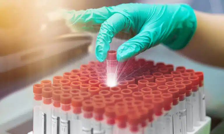
PBMC Isolation, Quality, and Sourcing: A Practical Guide for Researchers
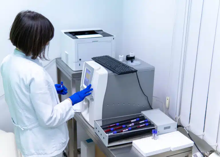
PBMC Isolation, Quality, and Sourcing: A Practical Guide for Researchers
Peripheral blood mononuclear cells — PBMCs — are the workhorse of human immunology research. They sit at the center of T cell assays, cytokine panels, drug screens, vaccine immunogenicity studies, and immune biomarker programs worldwide.
But any experiment built on PBMC cells is only as good as the underlying preparation. Quality depends on how the peripheral blood mononuclear cells were isolated, processed, cryopreserved, and characterized before they reached your bench. In practice, PBMC quality is one of the most variable and underappreciated drivers of assay reproducibility in the field.
This guide covers PBMC isolation methods, the quality variables that matter most, how to evaluate a preparation before you commit it to a study, and how to choose between fresh and cryopreserved formats. For foundational background, see What Are PBMCs? A Guide to Human Peripheral Blood Mononuclear Cells.
How Are PBMCs Isolated? The Density Gradient Method Explained
The standard method for isolating peripheral blood mononuclear cells from whole blood is density gradient centrifugation. It most often uses a polysucrose-based medium such as Ficoll-Paque, with a density of about 1.077 g/mL.
That density sits between the buoyant densities of mononuclear cells (~1.065–1.077 g/mL) and the denser granulocytes and red blood cells (>1.080 g/mL). This lets them separate in a single centrifugation step.
The isolation process runs as follows:
- Blood collection and dilution. Blood is collected into anticoagulant tubes (EDTA or heparin) and diluted with PBS or RPMI, usually 1:1. Dilution lowers viscosity and sharpens the density interface.
- Layering over density gradient medium. Diluted blood is carefully layered over Ficoll-Paque without disturbing the interface. This manual step is critical — technique affects how clean the separation is.
- Centrifugation. The tube is spun at about 400–500 × g for 30–40 minutes at room temperature, with no brake. Cells sediment by buoyant density. The brake setting matters: applying it disrupts the interface and contaminates the PBMC layer with granulocytes and red blood cells.
- Harvest of the buffy coat. The mononuclear cell layer forms a visible white ring at the blood-Ficoll interface. This buffy coat is carefully aspirated with a pipette and moved to a fresh tube.
- Washing. The harvested PBMC cells are washed two to three times in PBS or medium to remove residual Ficoll, platelets, and red blood cells. Each wash balances purity against centrifugation-induced cell stress.
- Counting and quality assessment. The final PBMC pellet is resuspended, counted, and checked for viability before use or cryopreservation.
Key fact: The whole PBMC isolation process, from blood collection to final resuspension, should finish within 4–6 hours of the blood draw. Each extra hour of delay is linked to measurable increases in monocyte activation, T cell stress marker expression, and viability loss.
What Affects PBMC Quality? The Six Critical Variables
The quality of a PBMC preparation reflects many variables across collection and processing. Whether you source peripheral blood mononuclear cells from a supplier or run your own isolation, understand each one.
1. Time from Blood Draw to Processing
This is the single most impactful pre-analytical variable in PBMC quality. Blood held at room temperature for more than 6–8 hours before processing undergoes progressive immune cell activation.
Monocytes raise IL-6 and TNF-α production thresholds. T cells start expressing activation markers such as CD69 and CD25 at baseline. Granulocyte degranulation releases elastase and other proteases into the suspension. As a result, PBMC cells from blood with long hold times show elevated spontaneous cytokine production, compressed stimulation windows, and altered NK cytotoxicity — artifacts that can be mistaken for real biology.
2. Temperature During Processing and Shipping
Both heat and cold damage mononuclear cells. Temperatures above 37°C speed up cell death and nonspecific activation. Temperatures below 4°C — especially freezing of unfixed samples — lyse cells and trigger cold-induced activation in monocytes.
For fresh PBMC products, ambient shipping in validated insulated packaging is generally fine for overnight transit. Longer shipping windows require active temperature control.
3. Donor Health Status and Immune State
The donor’s peripheral blood immune composition at collection directly sets the PBMC subset makeup and functional baselines.
Active infections, recent vaccinations, chronic inflammation, immunosuppressive medications, and even strenuous exercise in the prior 24–48 hours all measurably shift the T cell activation state, monocyte cytokine thresholds, and NK activity of the preparation. Research-grade peripheral blood mononuclear cells should come from donors confirmed healthy and off relevant medications at collection.
4. Centrifugation Parameters
Speed, temperature, time, and brake settings during density gradient separation all affect purity and yield. Too much force shears cells and adds granulocyte contamination. Too little leaves red blood cells incompletely sedimented and blurs the interface.
Processing above room temperature (22–25°C is optimal for Ficoll) reduces separation efficiency. Spinning with the deceleration brake on disrupts the interface and mixes cell layers — a common, and entirely preventable, cause of granulocyte contamination.
5. Cryopreservation Protocol (for Frozen PBMCs)
For cryopreserved peripheral blood mononuclear cells, the freezing protocol is the most consequential quality variable. Optimal cryopreservation requires three things:
- A validated cryoprotectant, typically 10% DMSO in heat-inactivated human AB serum or a serum-free equivalent.
- Controlled-rate cooling at −1°C/minute from room temperature to −80°C, using a controlled-rate freezer or an isopropanol “Mr. Frosty” device.
- Long-term storage in the vapor phase of liquid nitrogen (<−150°C).
Deviations at any step cause ice crystal formation that damages cell membranes and reduces post-thaw viability and function. Common culprits are too-rapid cooling, too little DMSO, or storage in the liquid phase of nitrogen, where temperature fluctuates during tank refilling.
Critical point: Post-thaw trypan blue viability alone does not confirm PBMC cryopreservation quality. Cells can exclude trypan blue while carrying functional deficits — impaired T cell proliferation, reduced NK cytotoxicity, elevated spontaneous cytokine production — that only show up in functional assays. Always request functional validation data alongside viability specs when evaluating a cryopreserved PBMC supplier.
6. Post-Thaw Recovery Protocol
For cryopreserved PBMC cells, the thaw protocol you use matters as much as the freeze protocol the supplier used.
Standard practice restores function: rapid thaw in a 37°C water bath (until a small ice crystal remains), immediate dilution into a large volume of pre-warmed medium to dilute the DMSO, and a short rest culture before assay setup (usually 2–4 hours at 37°C, 5% CO₂). This improves post-thaw recovery compared with slower thaw or immediate assay use.
SanguineBio’s human PBMCs are isolated from healthy, screened donors using validated density gradient protocols, processed within defined time windows and viability-tested at release, in both fresh and cryopreserved formats.
Fresh vs. Cryopreserved PBMCs: A Decision Framework
Both fresh and cryopreserved peripheral blood mononuclear cells work for research. The right choice depends on your application, logistics, and the functional properties you need to preserve.
| Consideration | Fresh PBMCs | Cryopreserved PBMCs |
|---|---|---|
| Monocyte function | ✅ Optimal — monocytes sensitive to freeze-thaw | ⚠️ Reduced recovery and function post-thaw |
| NK cell cytotoxicity | ✅ Maximal — no freeze-thaw effect | ⚠️ Reduced immediately post-thaw; recovers with rest culture |
| T cell proliferation assays | ✅ Preferred | ✅ Acceptable with quality cryopreservation |
| Antigen-specific T cell recall | ✅ Preferred | ✅ Well-validated in ELISpot and ICS assays |
| Longitudinal / multi-timepoint studies | ❌ Impractical | ✅ Required — banked aliquots thawed at each timepoint |
| Scheduling flexibility | ❌ Use within 24–48h of receipt | ✅ Thaw on demand |
| Multi-site assay standardization | ❌ Difficult to coordinate across labs | ✅ Single lot distributed to all sites |
| Cost per experiment | Higher (logistics-intensive) | Lower (flexible scheduling reduces waste) |
Quality Specifications to Require When Sourcing PBMCs
A research-grade PBMC preparation from a qualified supplier should meet these minimum specs. Evaluate any supplier who cannot document each one carefully before a critical study.
- Viability ≥90% for fresh, and ≥85% post-thaw for cryopreserved, measured by a nucleic acid dye (7-AAD or DAPI) rather than trypan blue alone, which overestimates viability in stressed mononuclear cells.
- Time-to-process documentation — the elapsed time from blood draw to completion of isolation, recorded and within a defined maximum (ideally <6 hours).
- Infectious disease screening — HIV-1/2, HBsAg, anti-HCV, HTLV-I/II, syphilis serology, and CMV serostatus, all tested at a CLIA-certified lab.
- Donor metadata — age range, sex, health status confirmed at collection, and a deidentified donor ID for lot-to-lot tracking and multi-lot studies from the same donor.
- Controlled-rate cryopreservation documentation (for frozen PBMC cells) — confirmation that controlled-rate freezing was used, not direct placement in −80°C freezers, which gives inconsistent cooling and inferior recovery.
- Platelet depletion — research-grade peripheral blood mononuclear cells should include platelet removal steps. Platelet contamination activates monocytes and generates false-positive innate cytokine signals.
When to Upgrade from PBMCs to a Leukopak
For most standard immunology assays, PBMC cells from a conventional blood draw are enough. But some applications outgrow what a standard PBMC preparation can provide:
- You need more than 500 million cells from a single donor to complete all experimental arms.
- You are developing a CAR-T manufacturing process and need to run multiple transduction conditions in parallel from one donor.
- You are running a large compound library screen in primary human immune cells and cannot tolerate inter-donor variability from pooling.
- You need billions of purified CD8+ T cells or CD4+ T cells for downstream genomics, CRISPR screening, or expansion protocols.
In these cases, a human leukopak is the right step up. It provides 5–30 billion mononuclear cells from a single leukapheresis collection. The cellular composition mirrors a standard PBMC preparation; only the scale changes.
Frequently Asked Questions About PBMC Isolation and Quality
What is the best method to isolate PBMCs?
Density gradient centrifugation over Ficoll-Paque (density 1.077 g/mL) is the gold-standard method for isolating peripheral blood mononuclear cells. It is highly reproducible when done within 6 hours of collection, at controlled temperature, with correct centrifugation parameters and no deceleration brake during the separation spin.
How long can PBMCs be stored at room temperature before processing?
PBMC quality begins to degrade measurably after 6–8 hours at room temperature, and optimal isolation is within 4 hours of the draw. Blood should never be refrigerated before processing. Cold accelerates platelet activation and monocyte stress that harm mononuclear cell quality.
How do I know if my cryopreserved PBMCs are good quality?
Post-thaw trypan blue viability is a starting point but not enough on its own. Functional validation gives the full picture: T cell proliferation to anti-CD3/CD28, NK cytotoxicity against K562 cells, and monocyte IL-6 production in response to LPS. Together these show whether the cryopreserved PBMC preparation is fit for downstream use.
What DMSO concentration is used to cryopreserve PBMCs?
The standard cryoprotectant for peripheral blood mononuclear cells is 10% DMSO in heat-inactivated human AB serum or a validated serum-free freezing medium. Below 10% gives incomplete cryoprotection. Above 10% adds cytotoxicity with no extra benefit.
Can PBMCs be isolated from leukopaks?
Yes. PBMC cells are routinely isolated from leukopaks using the same density gradient method applied to whole blood, scaled up for the larger starting volume. In this context, a leukopak is a high-yield source of mononuclear cells that allows PBMC isolation at scale from one characterized donor.
Summary: Key Facts About PBMC Isolation and Quality
- PBMCs are isolated by density gradient centrifugation over Ficoll-Paque — the gold-standard method that separates mononuclear cells from granulocytes and red blood cells in one step.
- The six critical quality variables are: time to processing, temperature, donor health status, centrifugation parameters, cryopreservation protocol, and post-thaw recovery.
- Blood should be processed within 4–6 hours of collection. Each extra hour of delay degrades PBMC functional quality measurably.
- Post-thaw trypan blue viability is not enough to confirm cryopreserved PBMC cell quality. Functional validation is required.
- Fresh peripheral blood mononuclear cells suit monocyte assays, NK cytotoxicity, and CAR-T transduction. Cryopreserved PBMCs suit longitudinal studies, multi-site standardization, and scheduling flexibility.
- When cell needs exceed a standard blood draw, leukopaks are the step up — billions of mononuclear cells from a single characterized donor.
Explore Sanguine’s human PBMC product page for current specifications and availability — isolated from healthy, screened donors using validated protocols, processed within defined time windows, and available in fresh and cryopreserved formats with full donor metadata.