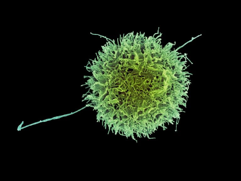
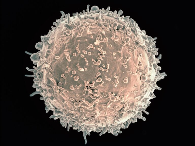
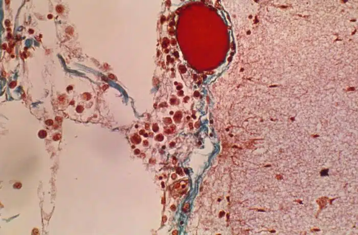
CD20-Negative Circulating Plasmablasts Are Target for New B Cell Therapies in Anti-CCP Positive RA
Diagnosing rheumatoid arthritis (RA) requires meeting several criteria set by the American College of Rheumatology and the European League Against Rheumatism. One criterion is the presence of anti-citrullinated protein antibodies (ACPA). Doctors currently test for ACPA using a filaggrin-derived peptide (anti-cyclic citrullinated peptide [anti-CCP]) ELISA. Other ELISAs in development, such as mutated citrullinated vimentin (MCV), show promising results.
Citrullination
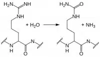
Myelin basic protein, filaggrin, and several histone proteins are naturally citrullinated. But other proteins, such as fibrin and vimentin, can become citrullinated during an inflammatory response. Citrullination is the enzymatic conversion of arginine residues into citrulline. It increases protein hydrophobicity, which can change the protein’s structure. In RA, immune cells recognize these citrullinated proteins as “non-self,” leading to ACPA production. Recent studies suggest these autoantibodies are not just convenient diagnostic markers left over from the autoimmune response — they may actually help drive RA. Current research in this area includes:
- Identifying RA subtypes based on ACPA positivity and specificity
- Determining how ACPA contributes to RA autoimmunity
Researchers know little about the B cells that produce ACPA. In a recent report in Annals of Rheumatic Disease, Kerkman and colleagues used B cells from the peripheral blood of ACPA-positive and ACPA-negative RA patients, plus healthy individuals, to study ACPA production in vitro and find out which cell populations produce it.
Testing ACPA Production in Isolated B Cells
The researchers first stimulated peripheral B cells with B cell activating factor (BAFF) and anti-IgM F(ab′)2-fragments to trigger ACPA production. Total IgG production was the same across the cultures, but only B cells from ACPA-positive RA patients produced ACPA. ACPA titers from in vitro culture matched in vivo patient ACPA titers, confirming that this model system works.
Next, the researchers looked at spontaneous ACPA production in unstimulated peripheral blood mononuclear cells (PBMC) from ACPA-positive RA patients. Here, total IgG was up to 100 times lower than in the stimulated B cell studies. But the amount of ACPA produced was about the same.
Identifying Which Cells Produce ACPA
Did circulating plasmablasts alone generate the ACPA in the PBMC cultures? Or did antigen presenting cells (APCs) in the PBMC population also push memory or even naïve B cells to make ACPA? Kerkman et al. used FACS to selectively deplete ACPA-positive RA patient PBMCs of plasmablast/plasma cell or naïve/memory populations, and to sort the CD19+ B cell subpopulations.
Results:
- Naïve B cells (CD20+CD27-) produced no ACPA at all, even when stimulated with BAFF and IgM F(ab′)2.
- Memory B cells (CD20+CD27+) produced ACPA upon stimulation, showing that CCP-specific memory cells circulate in ACPA-positive RA patients.
- However, ACPA production in CD20-depleted PBMCs stayed essentially unchanged, while unstimulated PBMCs depleted of plasmablasts/plasma cells produced much less ACPA.

What This Means for RA Treatment
This study shows that circulating ACPA-producing plasmablasts/plasma cells exist in the peripheral blood of ACPA-positive RA patients. This is a new and unexpected finding. Plasmablasts are normally a short-lived population within PBMCs after antigen exposure — antibody production usually continues from mature plasma cells in the spleen and lymph nodes instead.
Circulating ACPA-producing B cells may persist in RA for a few reasons: plasmablast replication, memory B cell activation in response to ongoing systemic citrullinated antigens, or both. Currently approved RA therapies that target the CD20+ B cell population, such as rituximab, affect memory B cells but not CD20- plasmablasts. New therapies that target circulating plasmablasts/plasma cells in addition to memory B cells could sharply limit ACPA production and the immune damage that follows in RA, including ACPA-induced TNFα production and complement activation. Identifying circulating plasmablasts as a major ACPA source is a step forward in understanding ACPA’s role in RA. It also points toward new therapies that target specific B cell populations in RA.
Further Reading:
Circulating plasmablasts/plasma cells as a source of anti-citrullinated protein antibodies in patients with rheumatoid arthritis. Kerkman PF, Rombouts Y, van der Voort EIH, Trouw LA, Huizinga TWJ, Toes REM, Scherer HU. Ann Rheum Dis 2013 Jul; 72:1259–1263.
The effect of targeted rheumatoid arthritis therapies on anti-citrullinated protein autoantibody levels and B cell responses. Modi S, Soejima M, Levesque MC. Clin Exp Immunol 2013 Jul; 173(1):8-17.
B effector cells in rheumatoid arthritis and experimental arthritis. Finnegan A, Ashaye S, Hamel KM. Autoimmunity 2012 Aug; 45(5):353-63.
Sanguine supplies research-grade human PBMCs for studies like this.