Unique anti-tumor functions of IFNg vs. IL-17 producing CD8 cells
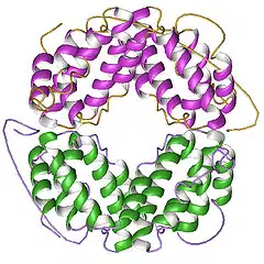
The anti-tumor power of IFNγ-producing CD4+ TH1 cells and CD8+ TC1 cells is well established. The role of IL-17 producing CD4+ (TH17) and CD8+ T cells (TC17) is less clear — different studies have found these cells can either inhibit or promote tumor growth. Context seems to be the key factor in whether IL-17-producing T cell subsets help or hurt anti-tumor immunity.
Comparing TC1 and TC17 Cells in a Melanoma Model
In the February 15, 2013 issue of The Journal of Immunology, Yu et al. compared how adoptively transferred tumor-specific TC1 and TC17 cells controlled tumor growth in mice. Here’s how the experiment worked:
- Researchers took anti-gp100 T cells from Pmel-1 TCR transgenic mice.
- They polarized these cells ex-vivo into either TC1 or TC17 phenotypes.
- They transferred the cells into mice with luciferase-expressing B16F10 melanoma lung metastases.
- These tumor-bearing mice had first undergone total body irradiation.
The team tracked tumor burden with luciferase luminescence and monitored overall survival. Both TC1 and TC17 cells fought the tumor, but TC1 cells did better: they stopped tumor growth completely, while TC17 cells only delayed it and eventually lost control of the tumor.
IFNγ Responsiveness Determines Which Cell Type Wins
The transferred TC1 cells made only IFNγ, not IL-17. TC17 cells made high levels of IL-17 and, as expected for this subset, could also turn into IFNγ-producing cells. The researchers found a striking result: removing IFNγ-responsiveness from tumor cells completely flipped which T cell type worked better.
- Tumor cells needed to respond to IFNγ for TC1 cells to fight them effectively — suggesting IFNγ-responsive genes help TC1 cells recognize tumor cells, slow their growth, or make them more prone to apoptosis.
- Once tumor cells could no longer respond to IFNγ, TC17 cells were able to drive complete tumor regression.
- Cytokine-neutralization tests showed IFNγ (not IL-17) was required in vivo for TC17 cells to work.
This suggests that in TC17 cell therapy, IFNγ needs to act on other cells in the tumor microenvironment — but IFNγ signaling inside the tumor cells themselves is, oddly, harmful to the therapy’s effect.
In the first few days after transfer, TC1 cells multiplied faster in vivo than TC17 cells. But by two and four weeks, TC17 cell levels in the spleen and lungs matched or exceeded TC1 levels — in mice with both wild-type and IFNγ-nonresponsive tumors.
This longer-term persistence may be part of why TC17 cells produce the anti-tumor responses they do.
The two pathways work differently: IFNγ signals through the STAT1 transcription factor, while IL-17 signals through a separate ACT1 pathway that activates NF-κB. TC17 cells also release IL-22, which activates STAT3. Crosstalk between these pathways inside tumor cells may explain why TC1 and TC17 cells depend so differently on IFNγ-responsiveness.
Implications for Adoptive Cell Therapy
It’s still not clear why TC17 cells in this model could control IFNγ-nonresponsive tumors but not wild-type tumors, while TC1 cells showed the opposite pattern. Still, these findings matter for how researchers design future adoptive cell transfer therapies.
Because TH17 cells persist long-term in vivo and show real anti-tumor activity, some researchers have proposed them as a strong CD4+ partner for CD8+ T cells in adoptive therapy. But this study suggests effectiveness will vary across cytokine-producing TC and TH subsets, depending on factors like whether the tumor can respond to cytokines such as IFNγ.
Further Reading
Adoptive Transfer of Tc1 or Tc17 Cells Elicits Antitumor Immunity against Established Melanoma through Distinct Mechanisms. Yu Y, Cho HI, Wang D, Kaosaard K, Anasetti C, Celis E, Yu XZ. J Immunol. 2013 Feb 15;190(4):1873-81.
Tumor-specific Th17-polarized cells eradicate large established melanoma. Muranski P, Boni A, Antony PA, Cassard L, Irvine KR, Kaiser A, Paulos CM, Palmer DC, Touloukian CE, Ptak K, Gattinoni L, Wrzesinski C, Hinrichs CS, Kerstann KW, Feigenbaum L, Chan CC, Restifo NP. Blood. 2008 Jul 15;112(2):362-73.
Phenotype, distribution, generation, and functional and clinical relevance of Th17 cells in the human tumor environments. Kryczek I, Banerjee M, Cheng P, Vatan L, Szeliga W, Wei S, Huang E, Finlayson E, Simeone D, Welling TH, Chang A, Coukos G, Liu R, Zou W. Blood. 2009 Aug 6;114(6):1141-9.
Structure and signalling in the IL-17 receptor superfamily. Sarah L. Gaffen. Nat Rev Immunol. 2009 August; 9(8): 556.
Human TH17 cells are long-lived effector memory cells. Kryczek I, Zhao E, Liu Y, Wang Y, Vatan L, Szeliga W, Moyer J, Klimczak A, Lange A, Zou W. Sci Transl Med. 2011 Oct 12;3(104):104ra100.
Photo credit: AJC1 via photopin cc