
Cell Therapy Manufacturing: Biospecimens for CAR-T and TCR Development
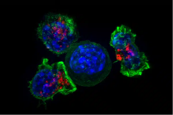
From Apheresis to Therapy: The Cell Manufacturing Journey
Chimeric antigen receptor T cell therapies have transformed how we treat blood cancers, a key application area within our oncology biospecimen portfolio. Six FDA-approved products show remarkable results in leukemia, lymphoma, and myeloma. Manufacturing starts with a patient blood collection and ends with cryopreserved cellular products.
The process turns blood cells into living therapies:
- Leukapheresis collections yield concentrated mononuclear cells
- T cell isolation follows
- Viral transduction introduces the CAR-encoding genes
- Ex vivo expansion grows the cells to a therapeutic dose
Each manufacturing step brings its own technical challenges, and quality control applies throughout. Regulatory rules govern every phase. Researchers work through these using well-characterized biospecimens from healthy donors and cancer patients.
Academic labs develop new CAR constructs using research-grade PBMCs. Phase I/II trials need increasingly rigorous documentation, while commercial manufacturing demands pharmaceutical-grade quality at scale.
Cellular therapy development moves from preclinical work through to commercial products, and each phase needs the right biospecimen quality level. From study design through receipt of samples, choosing the right material supports both regulatory compliance and scientific validity.
Leukopak: The Foundation of Cellular Immunotherapy
Leukopak units separate out mononuclear cells through automated apheresis. Continuous flow centrifugation concentrates lymphocytes, monocytes, and other PBMCs, while red blood cells and plasma go back to the donor.
Standard collections contain 5-20 × 10^9 total mononuclear cells, in volumes of 50-300 mL. That’s plenty of starting material for multiple manufacturing runs from a single donor unit.
The cellular makeup mirrors peripheral blood ratios:
- 70-80% lymphocytes, including 40-60% T cells, 10-25% B cells, and 5-15% NK cells
- 10-20% monocytes
- Variable granulocyte contamination, depending on collection parameters
Research Applications
For research, leukopak supports process development. Researchers use it to fine-tune T cell activation conditions and compare different activation bead formulations, including CD3/CD28 beads versus artificial antigen-presenting cells. It also supports:
- Testing lentiviral and retroviral vectors for viral transduction
- Formulating and testing expansion media
- Working out cryopreservation parameters that maximize post-thaw viability and function
Leukopak from cancer patients adds further value. It lets researchers assess how prior treatment affects T cell quality and test manufacturing feasibility across diverse patient populations. These include heavily pretreated, elderly, or immunocompromised patients, who present unique challenges.
This work surfaces patient-specific factors that predict manufacturing success or failure. Those insights then guide clinical trial enrollment criteria and patient selection strategies aimed at improving therapeutic success rates.
Purified T Cell Populations for CAR-T Development
Most CAR-T and TCR therapies specifically need T lymphocytes free of monocytes, B cells, NK cells, and other contaminants. Immunomagnetic selection using CD3 antibodies achieves this purification.
Positive Selection
Positive selection with anti-CD3 beads reaches over 95% CD3+ purity. Bead removal steps make sure no residual beads remain in the final product. Validation confirms any leftover bead presence won’t affect safety or efficacy.
Negative Selection
Negative selection depletes non-T cells using antibody cocktails. CD14 (monocytes), CD16 (NK cells, granulocytes), CD19 (B cells), and CD56 (NK cells) antibodies bind the unwanted cells. Magnetic bead separation removes them, leaving purified T cells behind.
Because negative selection doesn’t directly manipulate the T cells, it preserves their activation state. This benefits manufacturing processes that are sensitive to pre-activation, and some protocols perform better with this approach than with positive selection.
Further Fractionation
Further fractionation separates CD4+ helper T cells from CD8+ cytotoxic T lymphocytes. Defined CD4:CD8 ratios in the final product can improve persistence, reduce toxicity, or boost efficacy.
Selecting central memory T cells based on CD62L and CCR7 expression enriches for cells with stronger proliferative capacity. These cells keep more self-renewal potential, and CAR-T products made from central memory cells show better persistence in preclinical models.
Comparing bulk leukopak against purified T cells reveals real tradeoffs. Starting material cost, manufacturing complexity, product consistency, and quality attributes all need to be balanced. Some protocols use unselected leukopak to keep selection steps to a minimum. Others use defined T cell subset selection to optimize products for specific indications. Empirical testing determines the best strategy for a given CAR construct and tumor target.
Natural Killer Cell Therapies: Alternative Effector Cells
Natural killer cells are an attractive immunotherapy option:
- They recognize tumors without needing MHC matching
- They show potent natural cytotoxicity against stressed cells
- Allogeneic donors can be used without triggering graft-versus-host disease
- Their fast action kinetics suit aggressive cancers well, since they can intervene immediately without a lengthy manufacturing process
- Multiple dosing is feasible, unlike the single infusions typical of CAR-T
NK cell therapies in development include expanded autologous cells and allogeneic cells from healthy donors. Umbilical cord blood-derived NK cells offer an off-the-shelf option, and iPSC-derived NK cells provide an unlimited supply. CAR-NK constructs combine engineered targeting with natural cytotoxicity.
NK Cell Manufacturing
Manufacturing starts with leukopak starting material, followed by NK cell isolation. Anti-CD56 magnetic beads enable positive selection, while negative selection that depletes CD3+ T cells offers an alternative approach.
Isolated NK cell preparations then undergo ex vivo expansion. Cytokine cocktails with IL-2, IL-15, IL-21, and IL-12 drive proliferation. Feeder cells provide the costimulatory signals needed — often irradiated PBMCs, K562 cells, or engineered cell lines.
Expansion protocols lasting 14-21 days generate 100-1000 fold increases in cell numbers. Keeping cytotoxic function intact during expansion is a real challenge. Researchers have to work at preventing an exhausted phenotype from developing and carefully monitor for contaminating cell expansion.
CAR-NK Engineering
CAR-NK engineering adds further complexity. Viral transduction of primary NK cells is harder than T cell transduction. NK cells are less receptive to vectors and have a shorter ex vivo lifespan that limits expansion potential.
Alternative engineering approaches include:
- mRNA electroporation, which gives transient CAR expression
- Lentiviral vectors with modified envelopes that improve NK tropism
- Non-viral transposon systems that enable stable genomic integration
Researchers test these using purified NK cells from multiple donors.
T Cell Isolation and Activation Protocols
T cell purification from leukopak or PBMCs uses immunomagnetic selection. Positive selection with anti-CD3 beads captures T cells directly, while negative selection removes non-T cells using depletion antibodies.
The choice of selection method affects manufacturing performance. Positive selection reaches higher purity, over 98% CD3+, but the extra bead removal steps add complexity. Any residual beads must be validated below specification. Negative selection preserves more naïve activation states, since it doesn’t directly manipulate the T cells. Bead removal isn’t needed, because the beads bind to the cells being removed. Some evidence points to better expansion kinetics as a result.
Activation
Activation is the critical first manufacturing step. CD3/CD28 beads provide signal 1 (TCR) and signal 2 (costimulation). The bead-to-cell ratio affects how strong that activation is — higher ratios increase activation but risk over-stimulation.
Activation typically runs 24-72 hours. Adding IL-2 supports T cell survival and proliferation. Serum-free media formulations avoid animal-derived components for clinical manufacturing. Media composition has a major effect on both expansion and phenotype.
Transduction
Transduction happens during or shortly after activation, since activated T cells express surface receptors that make viral entry easier. Lentiviral vectors can transduce both resting and activated cells, while retroviral vectors need active cell division for genomic integration.
Transduction parameters include multiplicity of infection (MOI), transduction enhancers (polybrene, retronectin), and incubation time. Higher MOI increases transduction but raises vector costs and integration site complexity. Optimization means balancing efficiency against practical constraints.
Explore our full oncology biospecimen portfolio for additional cell therapy and cancer research sample types.


