Skin Punch Biopsy Samples for Research
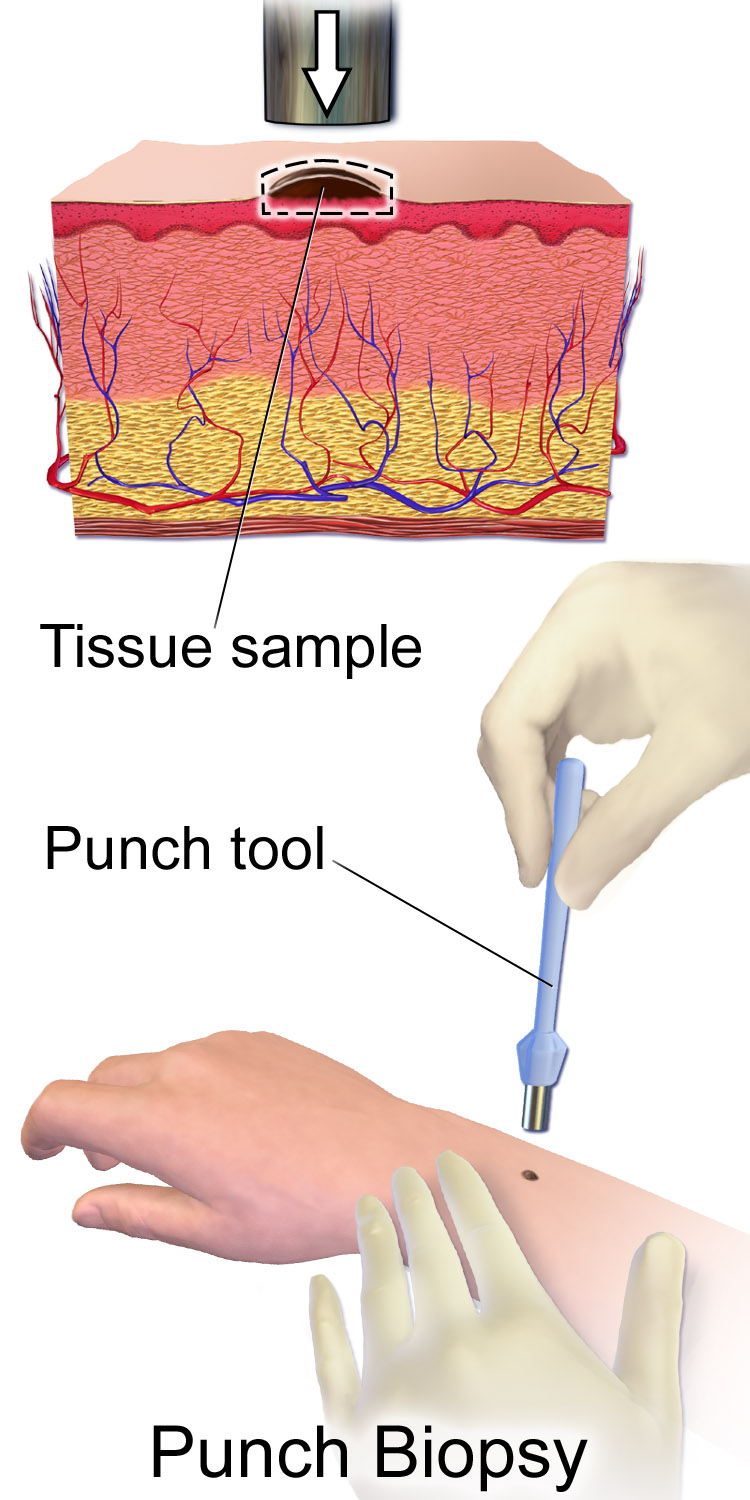
Skin is one of the few organs researchers can sample repeatedly, minimally invasively, and with the full architecture of the tissue intact. A skin punch biopsy is the standard way to do it — a small, cylindrical sample that captures every layer researchers typically need, from epidermis to subcutaneous fat.
What Is a Skin Punch Biopsy?
A punch biopsy uses a hollow, circular blade — typically 2mm to 8mm in diameter — to remove a cylindrical core of skin tissue. The tool is rotated through the epidermis and dermis and, depending on depth, into the subcutaneous fat, producing a full-thickness sample in a single, quick procedure. It’s the most commonly used skin sampling method in dermatology precisely because it’s fast, low-risk, and yields a architecturally complete sample.
What Does a Skin Punch Biopsy Sample Contain?
Depending on depth, a punch biopsy captures:
- Epidermis — the outer layer, including keratinocytes and resident Langerhans cells
- Dermis — connective tissue, blood vessels, nerve endings, hair follicles, and sweat glands
- Skin-resident immune cells — including dermal dendritic cells, tissue-resident memory T cells, and mast cells, which don’t circulate in peripheral blood and can only be studied directly in tissue
- Subcutaneous fat — if the punch is deep enough, providing access to adipose tissue and its associated stromal and immune populations
This makes punch biopsies uniquely valuable for research questions that blood-based sampling simply cannot answer.
Research Applications
- Dermatological disease research: Psoriasis, eczema, vitiligo, and other inflammatory or autoimmune skin conditions are studied directly in affected tissue, where disease-relevant immune infiltrates and structural changes are visible.
- Skin-resident immune cell studies: Tissue-resident memory T cells and dermal dendritic cells exist primarily in skin, not blood — punch biopsies are often the only way to access them.
- Histology and immunohistochemistry: Fixed, sectioned punch biopsies remain the standard for visualizing tissue architecture and marker expression in situ.
- Molecular and genomic profiling: RNA and DNA extracted from punch biopsies support gene expression studies, including single-cell and spatial transcriptomics of skin tissue.
- Wound healing and regenerative research: Punch biopsies themselves create a standardized wound, making the procedure both a sampling method and, in some study designs, a controlled injury model.
Fresh vs. Fixed or Frozen Processing
How a punch biopsy is processed determines what it can be used for. Fresh tissue, processed immediately, supports live-cell isolation and functional immune assays. Formalin-fixed, paraffin-embedded (FFPE) tissue is the standard for histology and immunohistochemistry, preserving structure at the cost of live-cell applications. Snap-frozen tissue preserves RNA and protein integrity for molecular assays while avoiding the fixation artifacts that can affect some antibody-based staining. The right processing path should be locked in before collection, since it isn’t reversible after the fact.
What to Evaluate When Sourcing Skin Punch Biopsies
- Diagnosis verification: For disease-state samples, confirm the donor’s diagnosis was clinically verified, not self-reported.
- Biopsy size and depth: Match the punch diameter and depth to your assay — deeper punches reach subcutaneous fat, but not every application needs it.
- Time to processing: Tissue viability for live-cell applications degrades with time; confirm the collection-to-processing window.
- Anatomical site: Skin biology varies meaningfully by body site (sun-exposed vs. non-exposed, lesional vs. non-lesional) — specify the site your study requires.
- Matched controls: For disease-state studies, request non-lesional skin from the same donor where possible, to control for inter-donor variability.
Frequently Asked Questions
How big is a skin punch biopsy sample?
Punch biopsies typically range from 2mm to 8mm in diameter, chosen based on the amount of tissue the research application requires and the acceptable level of donor discomfort.
Can skin punch biopsies capture immune cells?
Yes — skin harbors tissue-resident immune populations, including memory T cells and dendritic cells, that are not accessible from peripheral blood. This is one of the main reasons researchers use skin biopsies rather than blood-based sampling for certain immunology questions.
What’s the difference between a punch biopsy and a shave biopsy?
A punch biopsy removes a full-thickness cylindrical core, typically reaching the dermis or deeper. A shave biopsy removes only a thin, superficial slice and is generally not sufficient when full tissue depth or subcutaneous access is required.
Do skin punch biopsies need to be processed immediately?
For live-cell or functional applications, yes — processing time affects viability. For fixed histology or frozen molecular applications, biopsies are stabilized shortly after collection and can then follow standard storage timelines.
Explore Sanguine’s skin punch biopsy samples, available from healthy and disease-state donors with clinically verified diagnoses.